
December 19, 2008 (Milan, Italy) – For the first time, researchers have
shown that treating mild to moderate gum disease in otherwise healthy
volunteers improves endothelial dysfunction and significantly reduces
carotid intima media thickness (IMT), as measured by echo Doppler. The
report by Dr Stefania Piconi (Hospital Luigi Sacco, Milan, Italy) and
colleagues was published online December 12, 2008 in the FASEB Journal
[1].
“The novelty of this study is that this is the first physical evidence
that you can reverse a lesion that is already growing in the intima by
doing something as simple as taking care of your gums,” immunologist and
senior author Dr Mario Clerici (University of Milan, Italy) told
heartwire. “To tell you the truth, we were really surprised by the
result, but it turned up in subject after subject.”
http://www.medscape.com/viewarticle/585635
If you snore, your nightly noises may be a source of aggravation for the people around you, but snoring may actually be harmful to your health.
According to a story recently published in the journal Sleep, snoring is associated with a higher risk of carotid atherosclerosis, which is the accumulation of plaque in arteries that supply blood to the brain.
The researchers brought in 110 participants, including people who snored and others who didn’t. The participants underwent a sleep study, in which their snoring and breathing patterns were measured while they slept. They also underwent ultrasound assessment of their carotid arteries to measure atherosclerosis.
People who snored more often were much more likely to have carotid atherosclerosis, but curiously, not atherosclerosis in the arteries in their legs. One of the possible connections between snoring and this health risk is the vibration of the snoring. All that rattling in your throat may vibrate your carotid arteries, particularly a specific spot where plaque often forms. Researchers know that this vibration damages cells in artery walls, which could trigger the early formation of atherosclerosis.
This buildup of plaque can then set the stage for a stroke if a blood clot forms on the plaque blocking the artery, or if a piece of plaque breaks loose and becomes wedged in a smaller artery in your brain. According to the National Institutes of Health, more than half of all strokes in the United States are caused by plaque buildup in the carotid arteries, also known as carotid artery disease.
Methods for reducing snoring, as recommended by the National Sleep Foundation, include losing weight (this is the most effective step), sleeping on your side, avoiding alcohol several hours before bedtime, not smoking, and wearing a special device in your mouth that a dentist can prescribe.
Source: Lee, et al, Heavy Snoring as a Cause
of Carotid Artery Atherosclerosis, Sleep,
Sept. 1, 2008
The Missing Link Between Heart Disease and Dental Health
by HeartHawk
Sunday, January 04, 2009 For years there have been hints and hypotheses that heart disease and periodontal (gum) disease are associated or share common factors. Among the more humorous notions held by the uninformed press and public was that heart plaque and tooth plaque were somehow the same thing. That dubious notion notwithstanding there have long been provocative findings that have pointed to a relationship between a healthy mouth and a healthy heart.
The first and most widely studied theory was that the bacteria associated with periodontal disease (most notably Porphyromonas gingivalis) somehow traveled through the blood stream and provoked an inflammation response in the heart. Indeed, one of the first large studies (9760 participants over 17 years) found that “those with periodontitis had a 25% increased risk of coronary heart disease relative to those with minimal periodontal disease.” The link became even closer when a subsequent study determined that treating gum disease resulted in improved endothelial function and blood flow. Since then there have been numerous other studies that have detected a statistically significant association between gum disease and a variety of biomarkers for heart disease such as C-Reactive Protein (CRP) and Lipoprotein-associated Phospholipase A2 (Lp-PLA2).
One of the first direct links between periodontal and heart disease was found in a study that determined those with chronic periodontitis had higher triglyceride levels and a greater prevalence of small LDL a particularly powerful promoter of heart disease even among people with low cholesterol. The problem with these and many other studies is that it is often difficult to determine whether these similar biomarkers actually cause the disease or whether they are simply common indicators of a disease whose cause is some other common factor.
It could be that people without gum and/or heart disease simply live healthier, exercise, eat better, etc., than those with either or both diseases! However, for the first time, a study has shown that treating even mild gum disease in otherwise healthy people not only improves endothelial function but significantly reduces carotid intima media thickness (CIMT). That’s right, they found unequivocal evidence that treating gum disease regresses a standard measure of atherosclerosis.
To be fair, the study only looked at carotid arteries and not coronary arteries, it was a fairly small study (just 35 people), and CIMT is among these easiest markers of atherosclerotic lesions to regress. Head researcher Dr. Mario Clerici is quoted as stating, “The novelty ofthis study is that this is the first physical evidence that you can reverse a lesion that is already growing in the intima by doing something as simple as taking care of your gums . . . To tell you the truth, we were really surprised by the result, but it turned up in subject after subject.” The study involved nothing more than the simple removal of tartar and cleaning of the gums. There were no other procedures, no antibiotics or other prescription drugs or supplements, just the same basic dental hygiene measures you might receive at your dentist’s office.
Researchers used Echocardiography of carotid arteries to compare baseline CIMT against measurements made at several time points after treatment. They also measured common inflammatory biomarkers associated with cardiovascular risk. The study treatments resulted in significant reductions in CIMT at multiple sites as well as reductions in bacterial load and of the inflammation biomarkers. For the record, there is still much to be learned about the connection between heart disease and dental health. To recap the study was small, it only looked at carotid arteries not coronary arteries, and CIMT is perhaps the easiest atherosclerosis marker to regress. Nonetheless, we have the first solid evidence that there IS a connection between heart health and dental health. The takeaway heart health hint here is that you have another reason to follow the age-old admonition to visit your dentist regularly for a cleaning and check-up. You will now have two reasons to smile – whiter teeth and a potentially healthier heart.
he host’s reaction to foreign bodies known as an inflammatory response plays an important role in the etiology of cardiovascular disease. The physician is able to monitor this response by the serum levels of a blood protein called C-reactive protein or CRP. CRP levels in the blood have been shown to be predictive of a heart attack. In January of 2005 two papers were published that showed that if the blood levels of CRP were reduced, in these studies by prescription drugs known as statins, that the incidence of heart attacks were significantly decreased.
These studies indicate that the serum levels of CRP are an independent and modifiable risk factor for cardiovascular disease. But no one is quite sure what are the factors or conditions that cause the levels of CRP to increase in the blood. In most cases there are no acute infections or processes that can be associated with the elevated levels, leaving one to wonder what chronic, presumably asymptomatic condition is contributing to the high levels. In this context there is an emerging literature that suggests that dental infections, especially periodontal disease, could be playing a role. The connection is based on several observations that, while biologically plausible, lack the scientific rigor of a proven fact.
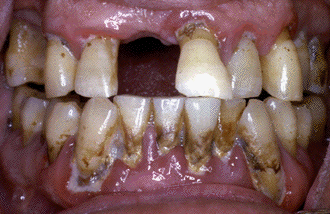
Could this man’s periodontal infection be a risk factor for cardiovascular disease?
In 1989 several cardiologists from Finland, i.e., Matilla, Syrjanen and their colleagues, reported that poor dental health could be associated with both an acute myocardial infarction and with a cerebral vascular accident. Subsequently, in a 7-year prospective study, dental disease as measured by the Total Dental Index (p=0.007), the number of previous myocardial infarctions (p=0.003), and to a lesser extent, diabetes (p=0.06), were associated with a risk of developing a new and often fatal myocardial infarction. Traditional risk factors, such as hypertension, smoking, total cholesterol levels, HDL cholesterol levels, triglycerides, social-economic status, gender and age were not significant predictors of a coronary event, when included in a model that contained the dental variables.
Other studies have generally confirmed this link between dental disease and coronary heart disease. A prospective cohort design study, involving data from 9,760 United States males examined three times between 1971 and 1987, found a significant relationship between either periodontitis or edentulism and coronary heart disease, even after adjusting for 13 known risk factors. A representative sample of 1,384 adult Finnish males, aged 45-64 years, showed that the number of missing teeth, along with hypertension, geographic area, and educational level were independent explanatory factors for the presence of ischemic heart disease. Among United States veterans participating in a longitudinal aging study, a significant association between periodontal disease, as measured by the extent of alveolar bone loss, and coronary heart disease and stroke could be demonstrated after adjusting for various cardiovascular risk factors.
We have been recording a large number of oral/dental variables in a group of elderly veterans, so as to study the relationship between oral/dental health and systemic diseases among older individuals. We found that a statistical association exists between a diagnosis of coronary heart disease and certain oral/dental parameters such as the numbers of missing teeth, plaque BANA test scores, salivary levels of certain bacteria and complaints of dry mouth or xerostomia. In logistic regression models, dentate individuals with 1 to 14 teeth were 2.81 times more likely to have coronary heart disease than individuals with most of their teeth, i.e., 15 to 28 teeth. A positive plaque BANA score, which indicates the presence of certain anaerobic bacteria in the plaque samples, was twice as likely to be found in dentate individuals with coronary heart disease, compared to dentate individuals without coronary heart disease. Individuals who complained of a dry mouth were 2.34 times more likely to have a diagnosis of coronary heart disease.
The dental/oral variables in these older individuals were more strongly associated with coronary heart disease than were recognized risk factors such as serum cholesterol levels, the body mass index and smoking status. This suggests that good dental health may be important in maintaining good cardiovascular health.
How could an inflammation about the gums cause disease on the linings of the arteries? Good question, as it is difficult to see how events occurring on the teeth or in the gum tissue could influence the development of an atheroma on the endothelial surface of arteries such as the coronary artery. A new paradigm is appearing in medicine that asks “are all diseases infections? This possibility was suggested by the demonstration that most ulcers are due to a treatable infection with Helicobacter pylori, and has been fueled in recent years by the association of chronic infections with cerebral and myocardial infarctions. Dental infections were not considered as contributory to these events, even though dental caries and periodontal disease are the most common of all chronic infections. It is well known that dental treatments, and dental infections can cause a bacteremia, and that this bacteremia has been associated with infective endocarditis.
Dental infections, involving the soft tissues of the periodontium and the pulp, can also elicit an inflammatory response that could release into the systemic circulation a variety of biologically active molecules. These bacterial products, such as lipopolysaccharides (LPS) and heat shock proteins (HSP), as well as inflammatory mediators such as cytokines, could directly or indirectly influence events on the intima of blood vessels. It is this propensity of dental infections to raise the white blood cell counts, to elicit a chronic inflammatory response, and/or an asymptomatic bacteremia, that will link dental disease with cardiovascular disease.
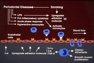
In the illustration shown below, smoking, which is a risk factor for both cardiovascular disease and periodontal infections, is shown to directly increase (upregulate) the number of adhesion molecules on the lining of endothelial cells. These adhesion molecules allow activated monocytes to attach to the lining of blood vessels and squeeze between the endothelial cells, causing an inflammatory response in the tissue below the endothelial cells. Smoking, however, promotes periodontal infections, which in turn releases LPS (lipopolysaccharide) molecules into the blood stream and these LPS molecules can also upregulate adhesion molecules. Other aspects of the inflammation in the gum tissues can also increase the blood levels of pro-inflammatory cytokines and acute phase proteins like the C-reactive proteins. The inflammation in the gum tissues can activate those monocytes which are capable of sticking to the adhesion molecules, which in turn can squeeze between the endothelial cells becoming tissue macrophages
Several chronic infections can follow this pathway of upregulating adhesion molecules and activating monocytes. The best known of these are respiratory infections due to Chlamydia pneumonia. Dental infections, however, are far more common than C. pneumonia infections and are usually asymptomatic. The man whose periodontal infection is shown below had no symptoms and came to the dental clinic seeking a replacement for his missing front tooth. He was oblivious to his periodontal condition, even though there was bleeding and inflammation around most of his teeth. Based upon recent studies, if we had taken a blood sample of this man, the levels of known risk factors for atherosclerosis (white blood cell count, C-reactive protein level, HDL-cholesterol level and fibrinogen level) would all be higher than those in a comparably aged man without periodontal disease. It is the extent and pervasiveness of the periodontal infection that would make this a possible risk factor for cardiovascular disease.

If periodontal inflammation is shown to be a risk factor for either coronary artery disease or stroke (see Dental Disease and Cerebral Vascular Disease), then it will be considered a modifiable risk factor, since it can be treated. This means that the dentist and the patient will both be concerned with periodontal disease and the available treatment choices. (See Antimicrobial Agents in Periodontal Disease).
from Walter Loesche: http://www.dent.umich.edu/research/loeschelabs/
Anaerobe. 2010 Dec;16(6):629-32. Epub 2010 Sep 8.
Presence of periodontopathic bacteria in coronary arteries from patients with chronic periodontitis.
Marcelino SL, Gaetti-Jardim E Jr, Nakano V, Canônico LA, Nunes FD, Lotufo RF, Pustiglioni FE, Romito GA, Avila-Campos MJ.
Department of Stomatology, University of São Paulo, SP, Brazil.
Abstract
In this study the presence of periodontopathic pathogens in atheromatous plaques removed from coronary arteries of patients with chronic periodontitis and periodontally healthy subjects by PCR was detected. Our results indicate a significant association between the presence of Porphyromonas gingivalis and atheromas, and the periodontal bacteria in oral biofilm may find a way to reach arteries.
JOURNAL OF MAXILLOFACIAL AND ORAL SURGERY Volume 8, Number 2, 108-113, DOI: 10.1007/s12663-009-0028-5
RESEARCH PAPER
Prevalence of periodontal pathogens in coronary atherosclerotic plaque of patients undergoing coronary artery bypass graft surgery
Jaideep Mahendra, Little Mahendra, V. M. Kurian, K. Jaishankar and R. Mythilli
Abstract
Background
Chronic bacterial infections have been associated with an increased risk for atherosclerosis and coronary artery disease. The ability of oral pathogens to colonize in coronary atheromatous plaque is well known. The aim of our study was to detect the presence of four common periodontal pathogens in coronary plaques. We detected the presence of 16S rRNA of Treponema denticola, Eikenella Corrodens, Porphyromonas gingivalis and Campylobacter rectus in subgingival and atherosclerotic plaques of CABG surgery by using Polymerase Chain Reaction.
Methods
51 patients in the age group of 40 to 80 years with chronic periodontitis were recruited for the study. These patients were suffering from Coronary Artery Disease (CAD) and underwent Coronary Artery Bypass Grafting (CABG). DNA was extracted from the subgingival plaque and coronary atheromatous plaque samples. Universal Primer for the general detection of bacterial DNA and the primers for T.denticola, E. Corrodens, C.rectus and P.gingivalis were used to amplify part of 16SrRNA gene by Polymerase Chain Reaction.
Results
T.denticola, E.corrodens, C.rectus and P.gingivalis were detected in 49.01 %, 27.45 %, 21.51% and 45.10% of atherosclerotic plaque samples. In both subgingival and coronary plaque samples, T. denticola was detected in 39.21% of the cases, E.corrodens in 19.60%, C.rectus in 11.76% and P.gingivalis in 39.22% of the cases respectively.
Conclusion
Our study revealed the presence of significant bacterial DNA of oral pathogens in coronary plaques. This suggests possible relationship between periodontal infection and atherosclerosis and can help devise preventive treatment strategies.
Total numbers of oral pathogens found to affect heart health more than a single type of bacteria
Posted on 2009-04-02 10:12:42 in Bone and Dental | Cardio-Vascular | Longevity and Age Management |
Researchers from the University of Buffalo Department of Oral Biology in the School of Dental Medicine conducted a study involving 386 men and women between the ages of 35 and 69, each of whom had suffered a heart attack. The study also included 840 heart-healthy individuals as controls. Oelisoa M. Andriankaja, D.D.S., Ph.D. and her colleagues collected samples of dental plaque – where germs adhere – from 12 different sites in the gums of all participants. They then analyzed the samples to determine the presence of six common forms of periodontal bacteria and to assess the total numbers of bacteria.
The participants who had suffered heart attacks were shown to have more of each type of bacteria than the control group. In addition, two species – Tannerella Forsynthesis and Preventella Intermedia – had a “statistically significant association with an increased risk of heart attack.” Results also showed that an increase in the number of different types of periodontal bacteria increased the odds of having a heart attack. “The message here is that even though some specific periodontal pathogens have been found to be associated with an increased risk of coronary heart disease, the total bacterial pathogenic burden is more important than the type of bacteria,” says Dr. Andriankaja. “In other words, the total number of ‘bugs’ is more important than one single organism.”
Dr. Andriankaja also noted that prospective studies that measure oral bacteria in participants who have no history of heart problems, but later suffer a heart attack, is needed to better assess the correlation between organisms that cause gum disease and the development of heart disease.
Levels of CRP may be a stronger predictor of potential heart attack or stroke than cholesterol, according to a study published in the Nov. 14, 2003 issue of the New England Journal of Medicine.
- Researchers have found that levels of both cholesterol and C-reactive protein were indicative of potential heart attacks and strokes, although the latter was more so. Also, women with high levels of one didn’t necessarily have high levels of the other.
- University of California Davis study identifies C-reactive protein as cause of blood clot formation. (Jan. 25, 2003 print edition of the journal Circulation – a publication of the American Heart Association). “The study provides further conclusive evidence that CRP, until now viewed as an ‘innocent bystander’ in the formation of heart disease, is in fact a key culprit that causes inflammation in the arteries, resulting in formation of clots and plaque that lead to heart attacks and strokes.
- Treatment of Periodontal Disease significantly reduces CRP levels. Okayama University Graduate School of Medicine and Dentistry, Okayama, Japan.
- Treating periodontal disease can significantly lower the levels of two inflammatory proteins associated with a heightened risk of heart disease, ,” Dr. Sara Grossi, senior author of the study, said. “Our results showed that in people who had elevated levels of CRP at baseline, removal of dental plaque bacteria by scaling or scaling combined with topical antibiotics produced a statistically significant reduction, bringing CRP levels close to the low-risk level. SUNY Buffalo.
-
Periodontal therapy lowers levels of heart disease inflammation markers Posted April 21, 2004 Treating periodontal disease with scaling and root planing combined with a topical antibiotic gel can significantly lower the levels of two inflammatory proteins associated with a heightened risk of heart disease, scientists from the State University of New York at Buffalo report.
Blood drawn from 102 subjects with periodontal disease showed elevated levels of both C-reactive protein and fibrinogen, proteins associated with increased risk for heart disease and blood clotting. All of the subjects were free of other conditions that could cause elevated levels of the proteins.
Scientists from the UB School of Dentistry’s Department of Oral Biology divided the subjects into two groups to determine if periodontal therapy would be effective in lowering the levels of the heart disease markers. One group received scaling and root planing treatment while the second group received treatment with the topical antibiotic Atridox followed by scaling and root planing.
Based on a treatment regimen at three, six and nine months and blood samples taken at six weeks and at three, six, nine and 12 months, repeated periodontal treatment resulted in a significant reduction in the systemic levels of the inflammation markers, the UB scientists said.
“People who have high levels of CRP in their blood are at high risk of heart disease,” Dr. Sara Grossi, senior author of the study, said. “Our results showed that in people who had elevated levels of CRP at baseline, removal of dental plaque bacteria by scaling or scaling combined with topical antibiotics produced a statistically significant reduction, bringing CRP levels close to the low-risk level.”
“Both treatments also significantly reduced levels of fibrinogen in patients with elevated fibrinogen levels,” she added.
Source: American Dental Association. http://www.ada.org/prof/resources/pubs/adanews/adanewsarticle.asp?articleid=841%20
Poor dental hygiene increases risk of heart attack and stroke
Not brushing your teeth may increase the risk of heart attack and stroke, according to the results of a recent study.
More than 700 different types of bacteria live in the mouth. Poor dental hygiene allows these bacteria to flourish, and bleeding gums gives them direct access to the bloodstream. Professor Howard Jenkinson, from the University of Bristol, England, and colleagues who led the study found that certain types of bacteria stick onto platelets causing the platelets to clump together and encase the bacteria, thus creating small blood clots.
The formation of these small blood clots increases a person’s risk of having a stroke or a heart attack. It also enables the bacteria to evade detection by the immune system and protects them from antibiotics, thus explaining why antibiotics do not always work when they are used to treat infectious heart disease.
“Cardiovascular disease is currently the biggest killer in the western world. Oral bacteria such as Streptococcus gordoniiand Streptococcus sanguinis are common infecting agents, and we now recognise that bacterial infections are an independent risk factor for heart diseases,” Said Professor Jenkins in a press release. “In other words it doesn’t matter how fit, slim or healthy you are, you’re adding to your chances of getting heart disease by having bad teeth.”
http://www.worldhealth.net/news/poor_dental_hygiene_increases_risk_of_he/
-
High Incidence of Actinobacillus Actinomycetemcomitans Infection in Acute Coronary Syndrome
Kaoru Sakurai1), Dongqing Wang2), Jun-ichi Suzuki1), Makoto Umeda2), Toshiyuki Nagasawa2), Yuichi Izumi2), Isao Ishikawa2)3) and Mitsuaki Isobe1)Recent epidemiological studies suggest that periodontitis is an important risk factor for coronary heart disease (CHD). The aim of this study was to evaluate the association between periodontitis and CHD, particularly acute coronary syndrome (ACS), focusing on microbiological and immunological features.
Twenty-eight CHD patients, 15 with ACS and 13 with chronic CHD, were included in this study. Coronary angiography, periodontal examination, and dental radiography were performed in all patients. Subgingival plaque, saliva, and blood samples were analyzed for the periodontopathogens Actinobacillus actinomycetemcomitans, Porphyromonas gingivalis, Tannerella forsythensis, Treponema denticola, and Prevotella intermedia using polymerase chain reaction.
Specific serum antibody titers to the 5 periodontal pathogens were determined by enzyme-linked immunosorbent assay. It was found that 33% of the ACS patients (5/15) harbored A. actinomycetemcomitans in oral samples, whereas no A. actinomycetemcomitans (0/13) was found in the chronic CHD patients (P < 0.05). Furthermore, ACS patients showed significantly higher serum IgG titers to A. actinomycetemcomitans (P < 0.05) compared with chronic CHD. More tooth loss and alveolar bone loss were noted in ACS patients than in chronic CHD patients, although the differences were not statistically significant.
Periodontal pathogens, particularly A. actinomycetemcomitans, may play a role in the development of ACS. -
Treatment of Periodontitis and Endothelial Function
New England Journal of Medicine Volume 356:911-920 March 1, 2007 Number 9 Maurizio S. Tonetti, D.M.D., Ph.D., Francesco D’Aiuto, D.M.D., Ph.D., Luigi Nibali, D.M.D., Ph.D., Ann Donald, Clare Storry, B.Sc., Mohamed Parkar, M.Phil., Jean Suvan, M.Sc., Aroon D. Hingorani, Ph.D., Patrick Vallance, M.D., and John Deanfield, M.B., B.Chir.
ABSTRACT
Background Systemic inflammation may impair vascular function, and epidemiologic data suggest a possible link between periodontitis and cardiovascular disease.
Methods We randomly assigned 120 patients with severe periodontitis to community-based periodontal care (59 patients) or intensive periodontal treatment (61). Endothelial function, as assessed by measurement of the diameter of the brachial artery during flow (flow-mediated dilatation), and inflammatory biomarkers and markers of coagulation and endothelial activation were evaluated before treatment and 1, 7, 30, 60, and 180 days after treatment.
Results Twenty-four hours after treatment, flow-mediated dilatation was significantly lower in the intensive-treatment group than in the control-treatment group (absolute difference, 1.4%; 95% confidence interval [CI], 0.5 to 2.3; P=0.002), and levels of C-reactive protein, interleukin-6, and the endothelial-activation markers soluble E-selectin and von Willebrand factor were significantly higher (P<0.05 for all comparisons). However, flow-mediated dilatation was greater and the plasma levels of soluble E-selectin were lower in the intensive-treatment group than in the control-treatment group 60 days after therapy (absolute difference in flow-mediated dilatation, 0.9%; 95% CI, 0.1 to 1.7; P=0.02) and 180 days after therapy (difference, 2.0%; 95% CI, 1.2 to 2.8; P<0.001). The degree of improvement was associated with improvement in measures of periodontal disease (r=0.29 by Spearman rank correlation, P=0.003). There were no serious adverse effects in either of the two groups, and no cardiovascular events occurred.
Conclusions Intensive periodontal treatment resulted in acute, short-term systemic inflammation and endothelial dysfunction. However, 6 months after therapy, the benefits in oral health were associated with improvement in endothelial function.
-
Ann N Y Acad Sci. 2006 Nov;1088:251-64
Low-grade inflammation in chronic infectious diseases: paradigm of periodontal infections. Moutsopoulos NM, Madianos PN.Oral Infection and Immunity Branch, National Institute of Dental and Craniofacial Research, National Institutes of Health, Bethesda, Maryland, USA.
Increasing evidence implicates periodontitis, a chronic inflammatory disease of the tooth-supporting structures, as a potential risk factor for increased morbidity or mortality for several systemic conditions including cardiovascular disease (atherosclerosis, heart attack, and stroke), pregnancy complications (spontaneous preterm birth [SPB]), and diabetes mellitus.
Cross-sectional, case-control, and cohort studies indicate that periodontitis may confer two- and up to sevenfold increase in the risk for cardiovascular disease and premature birth, respectively. Given the recently acquired knowledge that systemic inflammation may contribute in the pathogenesis of atherosclerosis and may predispose to premature birth, research in the field of periodontics has focused on the potential of this chronic low-grade inflammatory condition to contribute to the generation of a systemic inflammatory phenotype.
Consistent with this hypothesis clinical studies demonstrate that periodontitis patients have elevated markers of systemic inflammation, such as C-reactive protein (CRP), interleukin 6 (IL-6), haptoglobin, and fibrinogen. These are higher in periodontal patients with acute myocardial infarction (AMI) than in patients with AMI alone, supporting the notion that periodontal disease is an independent contributor to systemic inflammation. In the case of adverse pregnancy outcomes, studies on fetal cord blood from SBP babies indicate a strong in utero IgM antibody response specific to several oral periodontal pathogens, which induces an inflammatory response at the fetal-placental unit, leading to prematurity.
The importance of periodontal infections to systemic health is further strengthened by pilot intervention trials indicating thatperiodontal therapy may improve surrogate cardiovascular outcomes, such as endothelial function, and may reduce four- to fivefold the incidence of premature birth. Nevertheless, further research is needed to fully discern the underlying mechanisms by which local chronic infections can have an impact on systemic health, and in this endeavor periodontal disease may serve as an ideal disease model.
-
Atherosis: Volume 183, Issue 2, Pages 342-348 (December 2005)
21 of 29Associations between IgG antibody to oral organisms and carotid intima–medial thickness in community-dwelling adults
James D. Becka, Paul Ekeb, Dongming Linc, Phoebus Madianosd, David Coupere, Kevin Mossa, John Elterf, Gerardo Heissg, Steven Offenbacherc
Received 6 December 2004; received in revised form 25 February 2005; accepted 21 March 2005.
Abstract
Aims
The aims of this study are to describe the relationships between IgG antibodies to 17 oral organisms and atherosclerosis as indexed by carotid intima–medial wall thickness (IMT) and to evaluate the role of smoking.
Methods and results
Our study is based on a subset of participants in the Atherosclerosis Risk in Communities (ARIC) Study, who received a complete periodontal examination during visit 4 (1996–1998). The outcome was mean carotid IMT≥1mm assessed by B-mode ultrasound. The exposures were serum IgG antibody levels against 17 periodontal organisms using a whole bacterial checkerboard immunoblotting technique. Evaluation of all 17 antibodies indicated that antibody to Campylobacter rectus resulted in the best-fitting model (OR=2.3, 95% CI=1.83–2.84) and individuals with both high C. rectus and Peptostreptococcus micros titers had almost twice the prevalence of IMT≥1mm than those with only a high C. rectus antibody (8.3% versus 16.3%). Stratification by smoking indicated that all microbial models significant for smokers were also significant for never smokers except for Porphyromonas gingivalis (p=0.08).Conclusions
This is the first study to report a relationship between IgG antibody reactive to oral organisms and subclinical atherosclerosiswith significant relationships evident in both ever and never smokers. -
1: Eur J Vasc Endovasc Surg. 2007 Jul;34(1):102-6. Epub 2007 May 2.
Oral bacteria are a possible risk factor for valvular incompetence in primary varicose veins.
Kurihara N, Inoue Y, Iwai T, Sugano N, Umeda M, Huang Y, Ishikawa I.
Department of Vascular and Applied Surgery and Periodontology, Tokyo Medical and Dental University Graduate School of Medicine and Dentisty, Tokyo, Japan. [email protected]
OBJECTIVES: To investigate a possible link between valvular incompetence in primary varicose veins and chronic infection of periodontal disease by assessing the presence of oral bacteria in the great saphenous vein from patients with varicose veins and control subjects. MATERIAL AND METHODS: Forty-four primary varicose vein patients were enrolled in the study. 12 control saphenous veins were obtained from patients undergoing peripheral arterial bypass without clinical evidence of venous reflux. In total, 56 saphenous vein specimen (44 varicose veins and 12 control veins) were examined for 7 periodontal bacteria using a polymerase chain reaction (PCR) method. RESULTS: Of the 44 primary varicose vein patients, 31 patients were women and mean age was 59 years (range, 39-79 years). PCR examination of the diseased vein specimens showed that 48% were positive for at least one of 7 periodontal bacterial DNA. No bacteria were detected in the control specimens. CONCLUSION: Bacterial colonisation or infection of varicose veins is a frequent event although we were not able to establish whether this is a cause or consequence of the development of varices but this could be considered a risk factor for the development of varices.
-
Periodontitis May Increase Risk of Peripheral Arterial Disease
Chen YW, Umeda M, Nagasawa T et al. Eur J Vasc Endovasc Surg 2008; 35(2): 153-158.
A case controlled study looked at the association between periodontitis and PAD. 25 subjects having bypass surgery were matched with 32 healthy controls matched for age, sex, race and smoking history. DNA detection was used to look for periodontal pathogens. All subjects underwent a clinical periodontal examination and also had levels of IgG against the perio pathogens registered along with measures of their inflammatory cytokine levels.
Patients with PAD had a signifcantly higher prevalence of periodontitis and fewer teeth. DNA from the periodontal pathogens was detected in the arterial samples., higher IgG titers as well as significantly increased levels of the cytokines.
Regression analysis indicated that periodontis is associated with a five-fold increase in risk for PAD. The authors recognized that this does not confirm a causal relationship.
-
UC Davis study identifies C-reactive protein as cause of blood clot formation
(SACRAMENTO, Calif.) — Further underscoring the limitations of cholesterol screening in assessing a patient’s risk for heart disease, a new study by UC Davis physicians is the first to conclusively link C-reactive proteins (CRP) to formation of blood clots, a major cause of heart attacks, strokes and other vascular disease. Until now, CRP had been recognized mainly as a risk marker of heart disease. The study appears in the Jan. 25 print edition of the journal Circulation, a publication of the American Heart Association, and is available on the Web at www.circulationaha.org.
“The study provides further conclusive evidence that CRP, until now viewed as an ‘innocent bystander’ in the formation of heart disease, is in fact a key culprit that causes inflammation in the arteries, resulting in formation of clots and plaque that lead to heart attacks and strokes,” said Ishwarlal Jialal, professor of pathology and director of the Laboratory for Atherosclerosis and Metabolic Research at UC Davis School of Medicine and Medical Center.
The study demonstrates that CRP causes cells in the arteries, known as human aortic endothelial cells, to produce higher levels of an enzyme that inhibits the breakdown of clots. The enzyme, plasminogen activator inhibitor-1 (PAI-1) is also a strong risk marker for heart disease, especially in diabetics. The study used a variety of techniques to convincingly show how CRP activates PAI-1 in aortic cells, causing lesions in the arteries that ultimately lead to formation of plaque and blood clots.
The study underscores the need to use CRP screening to more accurately assess at-risk populations, according to Jialal, who is the Robert E. Stowell Endowed Chair in Experimental Pathology.
“Based on these findings, if a patient has normal cholesterol but high levels of CRP, an aggressive course of treatment is recommended to help the patient reduce the risk of heart attack, stroke and other heart diseases,” said Jialal. “By relying on cholesterol alone, a physician could significantly underestimate a patient’s risk level.” High CRP levels can occur in otherwise healthy individuals, according to the study. Patients with high levels of CRP can reduce risk by losing weight, exercising on a regular basis, stopping cigarette smoking, or taking statin drugs, Jialal added.
The study also closely links CRP and PAI-1 to diabetes and metabolic syndrome, a disorder characterized by a disproportionate amount of abdominal fat, elevated blood pressure, blood sugar and triglycerides and low levels of HDL, the “good” kind of cholesterol.
“In another important discovery, this study shows that in the presence of high blood-glucose levels, CRP is especially active in the stimulation of PAI-1. As a result, the effect of CRP is especially acute for patients with diabetes and metabolic syndrome,” said Sridevi Devaraj, a co-investigator and assistant professor of pathology at UC Davis. “Given the current pandemic of obesity which increases one’s risk of diabetes, the study’s insights about the active role of CRP and PAI-1 in heart disease are especially valuable.”
The new study adds to the findings of another landmark study on CRP by Jialal’s team at UC Davis that showed CRP actually damages the blood vessel wall by blocking a critical “protector” protein and inhibiting nitric oxide.
“Interestingly, the new study indicates that activation of PAI-1 was unrelated to the nitric oxide inhibition identified in the earlier study,” said Jialal. “This indicates that CRP has multiple, independent effects that cause heart disease.”
-

-
Porphyromonas gingivalis promotes murine abdominal aortic aneurysms via matrix metalloproteinase-2 induction
Aoyama N, Suzuki J, Wang D, Ogawa M, Kobayashi N, Hanatani T, Takeuchi Y, Izumi Y, Isobe M. Porphyromonas gingivalis promotes murine abdominal aortic aneurysms via matrix metalloproteinase-2 induction. J Periodont Res 2010; doi: 10.1111/j.1600-0765.2010.01326.x. © 2010 John Wiley & Sons A/S
Background and Objective: Abdominal aortic aneurysm (AAA) is a common and lethal disorder, and MMPs are highly expressed in AAA lesions. Large numbers of periodontopathic bacteria have been reported to be present in specimens obtained from the aortic walls of patients with an AAA. The purpose of this study was to analyze the influence of periodontopathic bacteria on AAA dilatation.
Material and Methods: AAAs were produced in mice by the periaortic application of 0.25 m CaCl2, and NaCl was used as a control. The mice were inoculated once weekly with live Porphyromonas gingivalis, live Aggregatibacter actinomycetemcomitans or vehicle.
Results: Four weeks after the periaortic application of either CaCl2 or NaCl, a significant increase was observed in the aortic diameter ofP. gingivalis-challenged mice compared with the vehicle control mice (p < 0.05), whereas there was no statistically significant increase in the aortic diameter of the A. actinomycetemcomitans-challenged mice. Immunohistochemical analysis found significantly higher numbers of CD8-positive and MOMA2-positive cells and significantly higher levels of MMP-2 in the aneurysmal samples of P. gingivalis-challenged mice compared with control mice. Live P. gingivalis promoted a significant proliferation of splenocytes in comparison with P. gingivalis-lipopolysaccharide and live A. actinomycetemcomitans (p < 0.05).
Conclusion: These findings demonstrate that challenge with P. gingivalis, but not with A. actinomycetemcomitans, can accelerate, or even initiate, the progression of experimental AAA through the increased expression of MMPs
Oral Microbiology and Immunology
Volume 24 Issue 1, Pages 64 – 68
-
Detection of oral bacteria in cardiovascular specimens
K. Nakano 1 , H. Nemoto 1 , R. Nomura 1 , H. Inaba 2 , H. Yoshioka 3 , K. Taniguchi 4 , A. Amano 2 , T. Ooshima 1
ABSTRACT
Background/aims: Oral bacteria, including cariogenic and periodontal pathogens, are thought to be etiological factors in the development of cardiovascular diseases. To define this relationship, we analyzed the distribution of oral bacterial species in cardiovascular specimens.
Method: Following acceptance into the study, 203 consecutive patients were analyzed, from whom 82 aortic valve specimens, 35 mitral valve specimens, and 86 aortic aneurysmal wall specimens, of which 16 contained aneurysmal thrombus tissues, were obtained. In addition, a total of 58 dental plaque specimens were collected from the same group of patients who underwent heart valve replacement or removal of aortic aneurysms. Bacterial DNA was extracted from both cardiovascular tissues and dental plaque in those cases and then species-specific polymerase chain reaction assays were used to analyze the occurrences of six oral streptococcal and six periodontal bacterial species.
Results: Streptococcus mutans was the most frequently detected species in the cardiovascular specimens, followed by Aggregatibacter actinomycetemcomitans. As for dental plaque specimens from patients who underwent cardiovascular operations, most of the tested periodontitis-related species as well as oral streptococci were detected at high frequencies. Furthermore, the positive rate of S. mutans in cardiovascular specimens from patients whose dental plaque specimens were also positive for S. mutans was 78%, which was significantly higher than any other tested species when the same analysis was performed.
Conclusion: Our results suggest that specific oral bacterial species, such as S. mutans and A. actinomycetemcomitans, are related to bacteremia and may be etiologic factors for the development of cardiovascular diseases.
-
JOURNAL OF MAXILLOFACIAL AND ORAL SURGERY
Volume 8, Number 2, 108-113, DOI: 10.1007/s12663-009-0028-5
RESEARCH PAPER
Prevalence of periodontal pathogens in coronary atherosclerotic plaque of patients undergoing coronary artery bypass graft surgery
Jaideep Mahendra, Little Mahendra, V. M. Kurian, K. Jaishankar and R. MythilliAbstract
Background
Chronic bacterial infections have been associated with an increased risk for atherosclerosis and coronary artery disease. The ability of oral pathogens to colonize in coronary atheromatous plaque is well known. The aim of our study was to detect the presence of four common periodontal pathogens in coronary plaques. We detected the presence of 16S rRNA of Treponema denticola, Eikenella Corrodens, Porphyromonas gingivalis and Campylobacter rectus in subgingival and atherosclerotic plaques of CABG surgery by using Polymerase Chain Reaction.
Methods
51 patients in the age group of 40 to 80 years with chronic periodontitis were recruited for the study. These patients were suffering from Coronary Artery Disease (CAD) and underwent Coronary Artery Bypass Grafting (CABG). DNA was extracted from the subgingival plaque and coronary atheromatous plaque samples. Universal Primer for the general detection of bacterial DNA and the primers for T.denticola, E. Corrodens, C.rectus and P.gingivalis were used to amplify part of 16SrRNA gene by Polymerase Chain Reaction.
Results
T.denticola, E.corrodens, C.rectus and P.gingivalis were detected in 49.01 %, 27.45 %, 21.51% and 45.10% of atherosclerotic plaque samples. In both subgingival and coronary plaque samples, T. denticola was detected in 39.21% of the cases, E.corrodens in 19.60%, C.rectus in 11.76% and P.gingivalis in 39.22% of the cases respectively.
Conclusion
Our study revealed the presence of significant bacterial DNA of oral pathogens in coronary plaques. This suggests possible relationship between periodontal infection and atherosclerosis and can help devise preventive treatment strategies. -
Systemic Immunoinflammatory Markers in Periodontitis Patients With Acute Myocardial Infarction A. URAZ1, G. TACOY1, M. KARCHED2, Z. TURGUT1, S. YUKSEL3, B. DOGAN4, A. BODUR1, and S. ASIKAINEN2, Objectives: Intensified proinflammatory reaction in arterial walls may render atheroma plaques prone to rupture and subsequent thrombus formation leading to cardiovascular complications, such as acute myocardial infarction (AMI). After gaining access to blood circulation components from periodontal bacteria may contribute as inflammatory triggers.
Here, the severity and systemic effects of periodontitis and a variety of systemic variables connected to cardiovascular risk were compared between periodontitis patients with or without a recent AMI. Methods: A total of 80 patients with periodontitis (mean age 50.3±6.7 years) were collected from Gazi University, Ankara; 40 of these periodontitis patients had been hospitalized with AMI (Per+AMI) and the remaining age- and gender-matched 40 periodontitis patients had no history of AMI (Per-AMI). In addition to demographic and life style variables, a full-mouth periodontal status was recorded and blood samples were taken on admission to hospital for inflammatory markers, lipid profile, glucose, and IgG levels against Porphyromonas gingivalis and Aggregatibacter actinomycetemcomitans outer membrane proteins (OMPs) and lipopolysaccharide (LPS). IgG levels were determined by ELISA. Results:
The CRP levels, white blood cell counts (WBC), glucose levels, BMI, total cholesterol, HDL-cholesterol, hypertension, smoking habits, dental plaque index, number of teeth, number of teeth with deep pockets, pocket depth, attachment level, and gingival bleeding values were significantly higher in Per+AMI than Per-AMI patients (p<0.01). IgG levels against A. actinomycetemcomitans LPS and OMP were significantly lower but those against P. gingivalis LPS and OMPs higher in periodontitis patients with than without AMI. Multiple logistic regression analysis identified the independent predictors, IgG levels against A. actinomycetemcomitans and P. gingivalis OMPs and LPS, CRP, total cholesterol, glucose levels, WBC, BMI, dental plaque, and gingival inflammation as risk factors for AMI. Conclusion:
The present result indicates that periodontitis infection may contribute to an increased risk for AMI.
–abstract from IADR, July 08
[Ed] So, this is the SECOND mechanism of attack. Initial inflammation/infection irritates the artery wall and begins the atheromatous plaque formation in cahoots with cholesterol.
THEN additional bouts of inflammation trigger a lysis process at the base of the plaque (using MMPs just like in periodontal attack) and loosen the plaque to the point that it detaches and becomes a floating clog or flapper valve occluding vessels locally or in other areas like the brain.
-

-
Mechanisms involved in the association between periodontal diseases and cardiovascular disease
- R Teles1,
- C-Y Wang3
Article first published online: 11 JAN 2011
DOI: 10.1111/j.1601-0825.2010.01784.x
© 2011 John Wiley & Sons A/S
Issue
- It is now well accepted that besides the cholesterol associated mechanisms of atherogenesis, inflammation plays a crucial role in all stages of the development of the atherosclerotic lesion. This ‘inflammation hypothesis’ raises the possibility that through systemic elevations of pro-inflammatory cytokines, periodontal diseases might also contribute to systemic inflammation and, therefore, to atherogenesis. In fact, there is evidence that periodontal diseases are associated with higher systemic levels of high-sensitivity C-reactive protein and a low grade systemic inflammation. This phenomenon has been explained based on mechanisms associated with either the infectious or the inflammatory nature of periodontal diseases. The purposes of this article were to review (1) the evidence suggesting a role for oral bacterial species, particularly periodontal pathogens, in atherogenesis; (2) the potential mechanisms explaining an etiological role for oral bacteria in atherosclerosis; (3) the evidence suggesting that periodontal infections are accompanied by a heightened state of systemic inflammation; (4) the potential sources of systemic inflammatory biomarkers associated with periodontal diseases; and (5) the effects of periodontal therapy on systemic inflammatory biomarkers and cardiovascular risk.
-
Bacteria Shown To Cause Blood Clots
Science News
04 Nov 2008
Bacteria can directly cause human blood and plasma to clot – a process that was previously thought to have been lost during the course of vertebrate evolution, according to new research at the University of Chicago, National Institute of Allergy and Infectious Diseases, and Institut Pasteur in Paris. Their findings were published online Nov. 2 in Nature Chemical Biology.
The discovery will improve scientists’ understanding of coagulation during bacterial infections and may lead to new clinical methods for treating serious medical conditions such as sepsis and anthrax.
It has long been known that blood often coagulates during sepsis or bacterial infections, but this has generally been regarded as a host’s immune and inflammatory response. It also has been known that bacteria can activate factors that precede coagulation, but it had not previously been known that bacteria can pass the coagulation threshold and cause blood clots to form. Once they form, the clots can grow and propagate. Although this may help prevent the dissemination of the bacteria through the host, it often leads to serious vascular damage due to blocked and injured blood vessels.
The key to clot formation is the location of the bacteria, rather than the total number of bacteria or their level of concentration. In other words, for those bacteria that can activate coagulation factors, coagulation occurs only when a cluster of bacteria forms.
“Our research demonstrates that coagulation can be controlled by changing the spatial distribution, or clustering, of bacteria,” said study co-author Christian Kastrup, Post-Doctoral Assistant at the Koch Institute for Integrative Cancer Research at the Massachusetts Institute of Technology. “Therefore, considering the location of bacterial cells, instead of just their presence or absence and their total numbers, could significantly change our understanding of coagulation.”
Kastrup, who worked on this research as a graduate student in the Ismagilov Lab at the University of Chicago’s Department of Chemistry, is the first author of the Nature paper. Rustem Ismagilov, Professor of Chemistry at the University of Chicago, is the corresponding author. Researchers at the National Institute of Allergy and Infectious Diseases, Institut Pasteur in Paris, and Ben-May Department for Cancer Research at the University of Chicago co-authored the paper.
Coagulation can occur if enough proteases that activate coagulation accumulate near the bacteria, rather than diffuse away. This research used Bacillus anthracis, the anthrax-causing pathogen (using a safe strain that does not infect humans). It found that in the case of human blood, coagulation required the secretion of zinc metalloprotease InhA1, which activated prothrombin and factor X directly – not via factor XII or tissue-factor pathways.
“We refer to this mechanism as ‘quorum acting’ to distinguish it from quorum sensing, in which bacteria coordinate certain actions based, in part, on their density,” said Wei-Jen Tang, Professor at the Ben-May Department for Cancer Research.
This work opens up a new field of st
udy, he added. “We will now explore the commonality of quorum acting, and how quorum acting can affect evolutionary dynamics.”The results of this research have broad implications, according to Ismagilov. “The work emphasizes the importance of bacteria’s spatial distribution, rather than just its average concentration in the functioning of nonlinear biochemical networks,” he said.


New research finds link between gum disease, acute heart attacks
By DAVID WILLIAMSON
UNC News ServicesCHAPEL HILL — Heart attack survivors who suffer advanced gum disease show significantly higher levels of a protein in their blood called C-reactive protein (CRP) than such patients without gum disease, new University of North Carolina at Chapel Hill research indicates.
The findings, presented Sunday (Nov. 12) during a news conference at the annual American Heart Association meeting in New Orleans, suggest that the presence of gum disease might increase the risk of a second heart attack in people with a history of heart disease.
“Not only did the heart attack patients with periodontal disease have higher levels of CRP than those without gum disease, but the CRP levels were directly related to the severity of the gum disease,” said Dr. Efthymios N. Deliargyris, an interventional cardiologist and a member of the Center for Oral and Systemic Diseases at UNC-CH. “The more severe the gum disease, the higher the CRP levels.”
Besides Deliargyris, also an instructor in medicine at the UNC-CH School of Medicine, study investigators were Drs. Steven Offenbacher, professor of periodontology and center director, James D. Beck, professor of dental ecology, both at the UNC-CH School of Dentistry, and Sidney C. Smith Jr., chief of cardiology and past president of the American Heart Association.
“We know a lot of risk factors for heart attacks, including high blood pressure, high cholesterol, diabetes and cigarette smoking, but all those combined only explain about two-thirds of heart attacks,” Deliargyris said. “Since about a third of people who suffer heart attacks don’t have those risk factors, there’s a wide search going on for other conditions that may contribute to increased risk.”
Studies at UNC-CH and elsewhere have linked periodontal disease — an advanced form of gingivitis — with increased risk of heart attacks, but it has been unclear what the two conditions have in common, the physician said.
“The one thing we know the two conditions share is that they tend to initiate an immune response, also called an inflammatory response, in the body,” he said. “The most common marker for this response is this C-reactive protein, which is considered predictive of future adverse events like heart attack.”
To learn how common severe gum disease was in heart attack victims, the UNC-CH team conducted their pilot study of 38 heart attack patients and matched them with a comparable group of 38 other people without known heart disease. Researchers found a high percentage of the former had periodontal disease — 85 percent — as compared with only 29 percent of the controls.
“The most exciting finding was that among people with a heart attack, those with periodontal disease had much higher CRP levels than those with a heart attack but no periodontal disease,” Deliargyris said. “It seems that the presence of periodontal disease on top of a heart attack has a synergistic effect and a very accentuated CRP release.”
Despite its small size, the study findings are the first of their kind and potentially very important, he said.
“This gives us an insight into possible mechanisms underlying the association between gum disease and heart disease,” Deliargyris said. “Now we believe that patients with a heart attack and periodontal disease have an exaggerated inflammatory response with higher CRP levels that might put them at risk for future heart attacks. This work also raises the possibility that by treating severe gum disease in people with heart attacks, we might be able to reduce their CRP levels and their risk of another heart attack.”
Journal of Thrombosis and Haemostasis doi:10.1111/j.1538-7836.2006.02128.x Infection with a periodontal pathogen induces procoagulant effects in human aortic endothelial cells G.A. Roth*, B. Moser*, S.J. Huang, J.S. Brandt, Y. Huang, P.N. Papapanou, A.M. Schmidt*, E. Lalla Summary Background: Multiple studies have demonstrated a link between periodontal infections and vascular disease. Porphyromonas gingivalis, a major periodontal pathogen, has been shown to adhere to and invade endothelial cells. Objective: In order to dissect mechanisms underlying these observations, we assessed the role of P. gingivalis infection in modulating properties of endothelial cells linked to atherothrombosis.
Methods: Primary human aortic endothelial cells (HAEC) were infected with either P. gingivalis 381 or its non-invasive fimbriae-deficient mutant, DPG3. Markers of coagulation and thrombosis were assessed 8h and 18h post-infection in cell lysates and supernatants. Results: Infection with P. gingivalis 381 significantly enhanced tissue factor expression and activity, and suppressed levels of tissue factor pathway inhibitor. Further, P. gingivalis infection decreased levels and activity of tissue plasminogen activator, and enhanced plasminogen activator inhibitor-1 antigen and activity. Consistent with an important role for bacterial adhesion/invasion in this setting, infection with DPG3 failed to induce procoagulant properties in HAEC. Most of the above effects of P. gingivalis 381 were more apparent at the later time point (18h post-infection). This suggests that P. gingivalis infection, rather than having an immediate and direct effect, might activate pathways that, in turn, trigger endothelial procoagulant mechanisms. Conclusions: Taken together these data demonstrate for the first time that infection with a periodontal pathogen induces procoagulant responses in HAEC.
Age-dependent associations between chronic periodontitis/edentulism and risk of coronary heart disease
Source: Circulation 2008 Apr1;117(13):1688-74 Authors: Dietrich T, Jimenez M, Krall Kaye EA, Vokonas PS, Garcia RI Summary of research: • Recognize the relationship between periodontitis and coronary heart disease (CHD), (angina, myocardial infarction or fatal CHD) in men.
• Significant association between periodontitis and CHD among men <60 years of age independent of diabetes, blood pressure, smoking, cholesterol etc.
• No such association found among men > 60 years of age.Results and conclusions: • Chronic periodontitis is associated with incidence of coronary heart disease among younger men (<60), independent of established cardiovascular risk factors. Key take-away • Male patients with chronic periodontal disease who are below the age of 60 have a higher risk of angina, heart attack or fatal coronary heart disease compared to men over the age of 60. Implementation strategies: • During routine risk assessment with all male patients under the age of 60 the clinician should make a statement (see sample below) regarding this research and comment about the importance of ongoing periodontal evaluation at each dental hygiene appointment.
Comparison of C-Reactive Protein and Low-Density Lipoprotein Cholesterol Levels in the Prediction of First Cardiovascular Events http://content.nejm.org/cgi/content/abstract/347/20/1557
Paul M. Ridker, M.D., Nader Rifai, Ph.D., Lynda Rose, M.S., Julie E. Buring, Sc.D., and Nancy R. Cook, Sc.D.ABSTRACT
Background Both C-reactive protein and low-density lipoprotein (LDL) cholesterol levels are elevated in persons at risk for cardiovascular events. However, population-based data directly comparing these two biologic markers are not available.
Methods C-reactive protein and LDL cholesterol were measured at base line in 27,939 apparently healthy American women, who were then followed for a mean of eight years for the occurrence of myocardial infarction, ischemic stroke, coronary revascularization, or death from cardiovascular causes. We assessed the value of these two measurements in predicting the risk of cardiovascular events in the study population.
Results Although C-reactive protein and LDL cholesterol were minimally correlated (r=0.08), base-line levels of each had a strong linear relation with the incidence of cardiovascular events. After adjustment for age, smoking status, the presence or absence of diabetes mellitus, categorical levels of blood pressure, and use or nonuse of hormone-replacement therapy, the relative risks of first cardiovascular events according to increasing quintiles of C-reactive protein, as compared with the women in the lowest quintile, were 1.4, 1.6, 2.0, and 2.3 (P<0.001), whereas the corresponding relative risks in increasing quintiles of LDL cholesterol, as compared with the lowest, were 0.9, 1.1, 1.3, and 1.5 (P<0.001). Similar effects were observed in separate analyses of each component of the composite end point and among users and nonusers of hormone-replacement therapy. Overall, 77 percent of all events occurred among women with LDL cholesterol levels below 160 mg per deciliter (4.14 mmol per liter), and 46 percent occurred among those with LDL cholesterol levels below 130 mg per deciliter (3.36 mmol per liter). By contrast, because C-reactive protein and LDL cholesterol measurements tended to identify different high-risk groups, screening for both biologic markers provided better prognostic information than screening for either alone. Independent effects were also observed for C-reactive protein in analyses adjusted for all components of the Framingham risk score.
Conclusions These data suggest that the C-reactive protein level is a stronger predictor of cardiovascular events than the LDL cholesterol level and that it adds prognostic information to that conveyed by the Framingham risk score.
Source InformationFrom the Center for Cardiovascular Disease Prevention and the Divisions of Preventive Medicine (P.M.R., L.R., J.E.B., N.R.C.) and Cardiology (P.M.R.), Brigham and Women’s Hospital and Harvard Medical School; and the Department of Laboratory Medicine, Children’s Hospital and Harvard Medical School (N.R.) — all in Boston.
-
Ann Thorac Surg 2004;77:537-543
© 2004 The Society of Thoracic SurgeonsRole of oral bacterial flora in calcific aortic stenosis: an animal model
David J. Cohen, MDa*, David Malave, MDa, John J. Ghidoni, MDb, Panagiotis Iakovidis, MDc, Mona M. Everett, PhDb,c,d, Shenghong You, MDd, Youhong Liu, MDd, Barbara D. Boyan, PhDd Abstract
BACKGROUND: Calcific aortic stenosis is a major public health problem in the United States. The mechanism of calcification remains unclear. The hypothesis that low grade chronic or recurrent bacterial endocarditis with specific calcifiable bacteria is a cause of calcification of the aortic valves was investigated using an animal model. Such bacteria are typically present as part of the normal human oral flora.METHODS: Forty New Zealand white rabbits were divided into four groups: group 1, control (1 ml of normal saline); group 2, Corynebacterium matruchotti 100,000 colonies; group 3, Streptococcus sanguis II 10 colonies; and group 4, C matruchotti 100,000 colonies plus S sanguis II 10 colonies. Animals were inoculated with bacteria through a flexible catheter placed through the aortic valve through a right carotid cut down. Inoculations were repeatedevery 3 days the first 2 weeks and then twice a week thereafter. At postmortem examination the aortic valves were harvested, embedded in paraffin, and stained with von Kossa stain. They were also examined by scanning and transmission electron micrography.RESULTS: Group 4 had 93.3% large calcifications (confluent calcium densities that are easily recognized with minimal magnification) and 6.6% small microcalcifications (dustlike microscopic particles requiring a compound microscope to appreciate) of the aortic valves. Group 3 exhibited large calcification in 20% and small in 40% of the aortic valves. Group 1 and group 2 had no evidence of calcification.
CONCLUSIONS: These results suggest that recurrent low-grade endocarditis from calcifying oral bacteria, particularly when occurring with synergistic strains, may be one cause of calcific aortic stenosis.
-
Abstract Journal of Periodontology 2007, Vol. 78, No. 2, Pages 322-327 (doi:10.1902/jop.2006.060081)
Evaluation of the Incidence of Periodontitis-Associated Bacteria in the Atherosclerotic Plaque of Coronary Blood Vessels Maciej Zaremba,* Renata Górska,* Piotr Suwalski, and Jan Kowalski*
Background: Unstable atherosclerotic plaque is a dangerous clinical condition, possibly leading to acute coronary deficiency resulting in cardiac infarction. Questions about the role of inflammatory factors in the formation of pathological lesions in the endothelium of coronary vessels have often been raised. This condition may be caused by bacteria that are able to initiate clot formation in a blood vessel, destabilizing an atherosclerotic plaque that is already present. The sources of these pathogens are chronic inflammatory processes occurring in the host, including periodontal disease, which is one of the most frequent conditions. The aim of this study was to evaluate the incidence of selected anaerobic bacteria in subgingival and atherosclerotic plaque in patients treated surgically because of coronary vessel obliteration.
Methods: The study was performed on 20 individuals with chronic periodontitis. Subgingival plaque was collected from periodontal pockets >5 mm. DNA testing was used to identify eight pathogens responsible for periodontal tissue destruction. Material from atherosclerotic plaques was collected from the same patients during bypass surgery, and DNA testing by the same method was performed.
Results: In 13 of 20 patients, the pathogens most frequently found in severe chronic periodontitis were also found in coronary vessels. In 10 cases, those species of bacteria were also present in atherosclerotic plaque. The most frequently identified bacteria werePorphyromonas gingivalis and Treponema denticola.
Conclusions: In patients with the severe form of chronic periodontitis, it seems that clinical attachment loss is not associated with bacterial permeability into coronary vessels. What is important is the presence of an active inflammatory process expressed by a significantly higher bleeding index in those patients in whom the examined bacterial species were found in atherosclerotic plaque.
-
Inflammation, heat shock proteins and periodontal pathogens in atherosclerosis: an immunohistologic study.
Ford PJ, Gemmell E, Chan A, Carter CL, Walker PJ, Bird PS, West MJ, Cullinan MP, Seymour GJ.
Oral Biology and Pathology, School of Dentistry, The University of Queensland, Brisbane, Australia. [email protected]
BACKGROUND: Inflammation is a significant component of atherosclerosis lesions. Bacteria, including periodontopathogens, have been demonstrated in atherosclerotic plaques and cross-reactivity of the immune response to bacterial GroEL with humanheat shock protein 60 has been suggested as a link between infections and atherosclerosis.
METHODS: In this study, the nature of the inflammatory infiltrate and the presence of human heat shock protein 60 and GroEL were examined in 31 carotid endarterectomy specimens. Additionally, monoclonal antibodies were used to detect the presence of six bacteria, including those implicated in periodontal disease. RESULTS: The inflammatory cell infiltrate of the lesions was dominated by CD14(+) macrophages and CD4(+) T cells. Most cells of the infiltrate as well as the endothelium were HLA-DR(+), indicating activation; however, there was an absence of CD25 expression, demonstrating that the activated T cells were not proliferating. Few CD1a(+) and CD83(+) cells were noted.
Human heat shock protein 60 expression was evident on endothelial cells and cells with the appearance of smooth muscle cells and lymphocytes. GroEL and bacteria were detected within intimal cells. Chlamydia pneumoniae, Porphyromonas gingivalis, Fusobacterium nucleatum, Tannerella forsythia, Prevotella intermedia, and Actinobacillus actinomycetemcomitans were found in 21%, 52%, 34%, 34%, 41%, and 17% of arteries, respectively. CONCLUSION: These results give evidence for a specific immune response associated with atherosclerosis. Whether bacteria initiate the observed inflammation in atherosclerotic lesions is not clear; however, the present study shows that maintenance of inflammation may be enhanced by the presence of periodontopathic bacteria.
Related Links
- Cross-reactivity of GroEL antibodies with human heat shock protein 60 and quantification of pathogens in atherosclerosis. [Oral Microbiol Immunol. 2005]
- Characterization of heat shock protein-specific T cells in atherosclerosis. [Clin Diagn Lab Immunol. 2005]
- Anti-P. gingivalis response correlates with atherosclerosis. [J Dent Res. 2007]
- Periodontal pathogens in atheromatous plaques. A controlled clinical and laboratory trial. [J Periodontal Res. 2004]
- Immunology of atherosclerosis. Demonstration of heat shock protein 60 expression and T lymphocytes bearing alpha/beta or gamma/delta receptor in human atherosclerotic lesions. [Am J Pathol. 1993]

Abstract 2007, Vol. 78, No. 4, Pages 677-682
(doi:10.1902/jop.2007.060062)
Correlation Between Atherosclerosis and Periodontal Putative Pathogenic Bacterial Infections in Coronary and Internal Mammary Arteries
Ana Pucar,* – Jelena Milasin,
–Vojislav Lekovic,* –Miroslav Vukadinovic,
–Miljko Ristic, ? –Svetozar Putnik,? and –E. Barrie Kenney
–
Background: Chronic infections, such as periodontitis, have been associated with an increased risk for atherosclerosis and coronary artery disease. The aim of this study was to investigate biopsy samples of coronary and internal mammary arteries for the presence of putative pathogenic bacteria (Porphyromonas gingivalis, Actinobacillus actinomycetemcomitans, Prevotella intermedia , and Tannerella forsythensis), Chlamydia pneumoniae, and human cytomegalovirus (CMV).
Methods: Patients with a diagnosis of coronary artery disease were included in the study. Fifteen coronary arteries with atherosclerosis and 15 internal mammary arteries without clinically assessable atherosclerotic degeneration were investigated. Both groups of specimens were obtained during coronary artery bypass grafting surgery. In all cases, the coronary and mammary artery specimens were taken from the same patient. The detection of periodontal pathogens, C. pneumoniae, and CMV was done by polymerase chain reaction analysis.
Results: Bacterial DNA was found in nine of 15 (60%) coronary artery biopsy samples: P. gingivalis in eight (53.33%), A. actinomycetemcomitans in four (26.67%), P. intermedia in five (33.33%), and T. forsythensis in two (13.33%) samples; CMV was detected in 10 (66.67%) samples, and C. pneumoniae was detected in five (33.33%) samples. Some of the samples contained more than one type of bacteria. Periodontal pathogens were not detected in internal mammary artery biopsies, whereas CMV was present in seven ( 46.67%) samples and C. pneumoniae was present in six (40%) samples.
Conclusion: The absence of putative pathogenic bacteria in internal mammary arteries, which are known to be affected rarely by atherosclerotic changes, and their presence in a high percentage of atherosclerotic coronary arteries support the concept that periodontal organisms are associated with the development and progression of atherosclerosis.
-
Periodontal Disease, C-Reactive Protein and Overall Health

Elevated C-reactive protein (CRP)levels increase the risk for cardiovascular disease Researchers have known for quite some time that elevated C-reactive protein (CRP) levels increase the risk for cardiovascular disease. A recent study published in The New England Journal of Medicine identified elevated CRP levels as a stronger predictor of heart attacks than elevated cholesterol levels, and recommended CRP and cholesterol screening for accurate risk assessment of cardiovascular disease.
However, many clinicians were unclear of the cause of elevated CRP levels. A study published earlier this year in the Journal of Periodontology reported that inflammatory effects from periodontal disease, a chronic bacterial infection of the gums, cause oral bacterial byproducts to enter the bloodstream and trigger the liver to make proteins such as CRP that inflame arteries and promote blood clot formation. Study Abstract
“Periodontal disease needs to be considered as a major contributor to increased levels of CRP by the medical community,” said Dr. Steven Offenbacher, member of the American Academy of Periodontology.
Previous studies reported that inflammatory effects from periodontal disease could cause oral bacterial byproducts to enter the bloodstream and trigger the liver to make proteins such as CRP that inflame arteries. In addition, these effects may cause blood clots that contribute to clogged arteries leading to heart attacks or strokes.
“What makes the recent findings noteworthy is that oral examinations were conducted on more than 5,000 adults in four U.S. communities already participating in a study to determine the risk of atherosclerosis,” said Offenbacher. “This is most likely the largest study confirming that periodontal disease and body mass index are jointly associated with increased levels of CRP in healthy adults.”
He added, “To reduce levels of CRP, and presumably the risk of cardiovascular disease, not only would it be important to lose weight if you are overweight, but it would also be important to get your gums treated.”
CRP testing is now available in many hospitals and health centers. The American Heart Association and the Centers for Disease Control and Prevention are developing a summary on whether CRP levels should be routinely tested to diagnose heart disease or to monitor progress of treatments.
“Based on this information and the potential to prevent heart attacks and strokes, I foresee patients receiving routine CRP testing in their dentist or periodontist office in the near future,” said Dr. Gordon Douglass, president of the American Academy of Periodontology. “This could help early diagnosis of potential heart disease sooner rather than later, as most people see their dentist or periodontist at minimum two times a year.”
source: http://www.perio.org/consumer/happy-heart.htm
-
http://www.sciencedaily.com/releases/2004/04 Repeated Treatment Of Gum Disease Reduces Levels Of Inflammatory Factors Known To Increase Heart Disease Risk
ScienceDaily (Apr. 8, 2004) — BUFFALO, N.Y. — Reinforcing the relationship between periodontal disease and heart disease, oral biologists from the University at Buffalo have shown that levels of two inflammatory proteins known to raise the risk of heart disease can be reduced substantially by regularly treating existing gum infections.
“You can have significant effects in other parts of the body by treating local problems,” said Sara Grossi, D.D.S., senior research scientist in the Department of Oral Biology, UB School of Dental Medicine and senior author on the study. “That’s why treating these infections is so important.”
Results of the study were presented at the International Association for Dental Research meeting held March 10-13 in Hawaii by Owais A. Farooqi, a doctoral student working with Grossi in UB’s Periodontal Disease Research Center.
The current study reports findings from 102 patients with periodontal disease who were randomized to two study groups and followed for one year. One group received standard “mechanical” treatment for periodontal disease, called scaling and root planning. The other group had the antibiotic gel Atridox™ applied to their gums before mechanical treatment.
Blood drawn at the start of the study showed that all patients had high levels of an inflammatory marker called C-reactive protein, which is known to put individuals at high risk for heart disease, and of fibrinogen, a protein involved in promoting blood clots. All patients were free of other conditions that could cause inflammatory proteins to show up in their blood stream
Both groups received their designated treatment at baseline, 3, 6 and 9 months. Blood samples were taken at baseline, and at 6 weeks, 3, 6, 9 and 12 months. At the end of 12 months, results showed that systemic levels of markers of inflammation decreased with repeated treatment.
“People who have high levels of CRP in their blood are at high risk of heart disease,” Grossi said. “Our results showed that in people who had elevated levels of CRP at baseline, removal of dental plaque bacteria by scaling or scaling combined with topical antibiotics produced a statistically significant reduction, bringing CRP levels close to the low-risk level. Both treatments also significantly reduced levels of fibrinogen in patients with elevated fibrinogen levels.
Grossi’s lab next will assess levels of cytokines — additional markers of inflammation — and how periodontal therapy affects them. “This work will provide additional information on the systemic effect or benefit of treating gum infection and provide valuable information on the relationship between gum disease and heart disease and diabetes.
Additional researchers on the study were Alex Ho, statistician, and Robert J. Genco, D.D.S., Ph.D., SUNY Distinguished professor, from the UB Department of Oral Biology, and J. Steven Garrett from Atrix Laboratories Inc.
-
Prim Dent Care. 2007 Apr;14(2):59-66.
Cardiovascular and oral disease interactions: what is the evidence?
Ford PJ, Yamazaki K, Seymour GJ.
Oral Biology and Pathology, School of Dentistry, University of Queensland, Brisbane, Australia. [email protected]
This paper reviews the evidence for the interaction of oral disease (more specifically, periodontal infections) with cardiovascular disease. Cardiovascular disease is a major cause of death worldwide, with atherosclerosis as the underlying aetiology in the vast majority of cases. The importance of the role of infection and inflammation in atherosclerosis is now widely accepted, and there has been increasing awareness that immune responses are central to atherogenesis. Chronic inflammatory periodontal diseases are among the most common chronic infections, and a number of studies have shown an association between periodontal disease and an increased risk of stroke and coronary heart disease. Although it is recognised that large-scale intervention studies are required, pathogenic mechanism studies are nevertheless required so as to establish the biological rationale. In this context, a number of hypotheses have been put forward; these include common susceptibility, inflammation via increased circulating cytokines and inflammatory mediators, direct infection of the blood vessels, and the possibility of cross-reactivity or molecular mimicry between bacterial and self-antigens. In this latter hypothesis, the progression of atherosclerosis can be explained in terms of the immune response to bacterial heat shock proteins (HSPs). Because the immune system may not be able to differentiate between self-HSP and bacterial HSP, an immune response generated by the host directed at pathogenic HSP may result in an autoimmune response to similar sequences in the host. Furthermore, endothelial cells express HSPs in atherosclerosis, and cross-reactive T cells exist in the arteries and peripheral blood of patients with atherosclerosis. Each of these hypotheses is reviewed in light of current research. It is concluded that although atherosclerotic cardiovascular disease is almost certainly a multifactorial disease, there is now strong evidence that infection and inflammation are important risk factors. As the oral cavity is one potential source of infection, it is wise to try to ensure that any oral disease is minimised. This may be of significant benefit to cardiovascular health and enables members of the oral health team to contribute to their patients’ general health.
-
Periodontal Infections and Coronary Heart Disease
Role of Periodontal Bacteria and Importance of Total Pathogen Burden in the
Coronary Event and Periodontal Disease (CORODONT) Study
Axel Spahr, DDS; Elena Klein, DDS; Natalie Khuseyinova, MD; Clemens Boeckh,
PhD; Rainer Muche, PhD; Markus Kunze, MS; Dietrich Rothenbacher, MD, MPH; Gita
Pezeshki, PhD; Albrecht Hoffmeister, MD; Wolfgang Koenig, MDArch Intern Med. 2006;166:554-559.
Background Chronic inflammation from any source is associated with increased
cardiovascular risk. Periodontitis is a possible trigger of chronic
inflammation. We investigated the possible association between periodontitis
and coronary heart disease (CHD), focusing on microbiological aspects.
Methods A total of 789 subjects (263 patients with angiographically confirmed,
stable CHD and 526 population-based, age- and sex-matched controls without a
history of CHD) were included in the Coronary Event and Periodontal Disease
(CORODONT) study. Subgingival biofilm samples were analyzed for periodontal
pathogens Actinobacillus actinomycetemcomitans, Tannerella forsythensis,
Porphyromonas gingivalis, Prevotella intermedia, and Treponema denticola using
DNA-DNA hybridization. The need for periodontal treatment in each subject was
assessed using the Community Periodontal Index of Treatment Needs (CPITN). The
main outcome measures included total periodontal pathogen burden, number of the
various periodontal pathogens in the subgingival biofilm, and periodontal
treatment needs (according to the CPITN).Results In multivariable analyses, we found a statistically significant
association between the periodontal pathogen burden (log10 of the sum of all
pathogens) (odds ratio [OR], 1.92; 95% confidence interval [CI], 1.34-2.74;
P<.001) or the number of A actinomycetemcomitans in periodontal pockets (log10)
(OR, 2.70; 95% CI, 1.79-4.07; P<.001) and the presence of CHD. In addition, a
statistically significant association between an increase in mean CPITN score
by 1 and the presence of CHD (OR, 1.67; 95% CI, 1.08-2.58; P = .02) was
observed.Conclusions Our findings suggest an association between periodontitis and
presence of CHD. Periodontal pathogen burden, and particularly infection with A
actinomycetemcomitans, may be of special importance. -
Anders Holmlund, Gunnar Holm, Lars Lind. Severity of Periodontal Disease and Number of Remaining Teeth Are Related to the Prevalence of Myocardial Infarction and Hypertension in a Study Based on 4,254 Subjects. Journal of Periodontology 2006, Vol. 77, No. 7, Pages 1173-1178
Background: During the past 15 years, mounting evidence for the association between periodontal and cardiovascular disease has been presented in epidemiological studies. The aim of this study was to investigate how the severity of periodontal disease and number of remaining teeth relates to myocardial infarction (MI) and hypertension (HT).
Methods: Self-reported history of HT and MI was collected in 3,352 patients referred to the Department of Periodontology, Gävle County Hospital, and in 902 subjects randomly selected from the general population. Severity of periodontitis was estimated by a combination of the amount of bone loss around each tooth investigated from a full-mouth x-ray, the presence or absence of bleeding on probing (BOP), and involvement of furcations.
Results: The severity of periodontitis was significantly associated with HT (prevalence 16%; P <0.0005), even after adjustment for age, gender, number of teeth, and smoking in the total sample, and with MI (prevalence 1.7%, P <0.03) after above-mentioned adjustments, but in middle-aged (40 to 60 years) subjects only. The number of diseased periodontal pockets was related to HT only (P <0.0001), and this relationship remained after the above-mentioned adjustments. The number of teeth was associated with MI (P <0.03) even after correction for age, gender, and smoking but was not related to hypertension.
Conclusions: The severity of periodontal disease was related to HT independent of age but to the prevalence of MI in middle-aged subjects only. The number of diseased pockets was significantly related to HT only. On the other hand, the number of teeth was associated with the prevalence of MI independent of age but not to HT. These data support the view that oral health is related to cardiovascular disease in a dose-dependent manner.
-
First Study To Take Bacteria Directly From Patients’ Mouths Further Supports Possible Link Between Gum And Heart Disease
ScienceDaily (Feb. 27, 1998) — By taking bacteria samples directly from patients’ mouths and exposing the samples to human blood platelets, researchers at Temple Univeristy Schools of Dentistry and Medicine have further confirmed a possible link between periodontal bacteria and heart disease.
In recent weeks, a great deal of media attention has been focused on this possible relationship. In fact, since the early 1990s, preliminary studies at other universities have used laboratory strains of mouth bacteria and population data to show this possible link.
Temple’s study is the first to take a large number of dental plaque bacteria directly from the mouths of patients with severe periodontal disease and test their effect on blood platelets.
“Almost immediately after we exposed human blood platlets to the dental plaque bacteria, the platelets began to clump together,” says Dr. Eugene J. Whitaker, associate professor of Dentistry and lead investigator. “And, out of all the periodontal bacteria we tested, Porphyromanas gingivalis was the only one to cause this clumping, which is a key step in formation of bloodstream thrombi (blockage).”
Porphyromanas gingivalis is the most important bacterial cause of destructive gum diseases in adults. The Temple research findings further support and expand a possible link between periodontal disease and development of athrosclerotic heart disease, a condition resulting from plaque build-up and constriction of coronary heart arteries, and strokes affecting the brain.
“The importance of our findings is that at least 36 million American adults have some form of destructive periodontal disease, which leads to loosening and loss of teeth,” says Dr. Thomas E. Rams, a co-investigator and chairman of Temple’s Department of Periodontology. “Porphyromanas gingivalis is very frequently in dental plaque causing this disease. These people may be at increased risk of getting heart disease and strokes if Porphyromanas gingivalis from their mouth gets into the bloodstream and clumps platelets similar to what we see in the laboratory.”
Note the date is 1998!–Ed
-
http://www.sciencedaily.com/releases/2000/11/ 001113071724.htm New Research Finds Link Between Gum Disease, Acute Heart Attacks
ScienceDaily (Nov. 13, 2000) — CHAPEL HILL — Heart attack survivors who suffer advanced gum disease show significantly higher levels of a protein in their blood called C-reactive protein (CRP) than such patients without gum disease, new University of North Carolina at Chapel Hill research indicates.
The findings, presented Sunday (Nov. 12) during a news conference at the annual American Heart Association meeting in New Orleans, suggest that the presence of gum disease might increase the risk of a second heart attack in people with a history of heart disease.
“Not only did the heart attack patients with periodontal disease have higher levels of CRP than those without gum disease, but the CRP levels were directly related to the severity of the gum disease,” said Dr. Efthymios N. Deliargyris, an interventional cardiologist and a member of the Center for Oral and Systemic Diseases at UNC-CH. “The more severe the gum disease, the higher the CRP levels.”
Besides Deliargyris, also an instructor in medicine at the UNC-CH School of Medicine, study investigators were Drs. Steven Offenbacher, professor of periodontology and center director, James D. Beck, professor of dental ecology, both at the UNC-CH School of Dentistry, and Sidney C. Smith Jr., chief of cardiology and past president of the American Heart Association.
“We know a lot of risk factors for heart attacks, including high blood pressure, high cholesterol, diabetes and cigarette smoking, but all those combined only explain about two-thirds of heart attacks,” Deliargyris said. “Since about a third of people who suffer heart attacks don’t have those risk factors, there’s a wide search going on for other conditions that may contribute to increased risk.”
Studies at UNC-CH and elsewhere have linked periodontal disease — an advanced form of gingivitis — with increased risk of heart attacks, but it has been unclear what the two conditions have in common, the physician said.
“The one thing we know the two conditions share is that they tend to initiate an immune response, also called an inflammatory response, in the body,” he said. “The most common marker for this response is this C-reactive protein, which is considered predictive of future adverse events like heart attack.” To learn how common severe gum disease was in heart attack victims, the UNC-CH team conducted their pilot study of 38 heart attack patients and matched them with a comparable group of 38 other people without known heart disease. Researchers found a high percentage of the former had periodontal disease — 85 percent — as compared with only 29 percent of the controls.
“The most exciting finding was that among people with a heart attack, those with periodontal disease had much higher CRP levels than those with a heart attack but no periodontal disease,” Deliargyris said. “It seems that the presence of periodontal disease on top of a heart attack has a synergistic effect and a very accentuated CRP release.”
Despite its small size, the study findings are the first of their kind and potentially very important, he said.
“This gives us an insight into possible mechanisms underlying the association between gum disease and heart disease,” Deliargyris said. “Now we believe that patients with a heart attack and periodontal disease have an exaggerated inflammatory response with higher CRP levels that might put them at risk for future heart attacks. This work also raises the possibility that by treating severe gum disease in people with heart attacks, we might be able to reduce their CRP levels and their risk of another heart attack.”
-
Curr Pharm Des. 2007;13(36):3665-75.
Porphyromonas gingivalis mediated periodontal disease and atherosclerosis: disparate diseases with commonalities in pathogenesis through TLRs.
Department of Medicine, Boston University School of Medicine, Boston, MA 02118, USA.
Toll-like receptors (TLRs) are a group of pathogen-associated molecular pattern receptors, which play an important role in innate immune signaling in response to microbial infection. It has been demonstrated that TLRs are differentially up regulated in response to microbial infection and chronic inflammatory diseases such as atherosclerosis. Furthermore hyperlipidemic mice deficient in TLR2, TLR4, and MyD88 signaling exhibit diminished inflammatory responses and decreased atherosclerosis. Accumulating evidence has implicated specific infectious agents including the periodontal disease pathogen Porphyromonas gingivalis in the progression of atherosclerosis. Evidence in humans suggesting that periodontal infection predisposes to atherosclerosis is derived from studies demonstrating that the periodontal pathogen P. gingivalis resides in the wall of atherosclerotic vessels and seroepidemiological studies demonstrating an association between pathogen-specific IgG antibodies and atherosclerosis. We have established that the inflammatory signaling pathways that P. gingivalis utilizes is dependent on the cell type and this specificity clearly influences innate immune signaling in the context of local and distant chronic inflammation induced by this pathogen. We have demonstrated that P. gingivalis requires TLR2 to induce oral inflammatory bone lose in mice. Furthermore, we have demonstrated that P. gingivalis infection accelerates atherosclerosis in hyperlipidemic mice with an associated increase in expression of TLR2 and TLR4 in atherosclerotic lesions. Our recent work with P. gingivalis has demonstrated the effectiveness of specific intervention strategies (immunization) in the prevention of pathogen-accelerated atherosclerosis. Improved understanding of the mechanisms driving infection, and chronic inflammation during atherosclerosis may ultimately provide new targets for therapy.
-
1: Circulation. 2004 Jun 8;109(22):2801-6. Epub 2004 May 3. Innate immune recognition of invasive bacteria accelerates atherosclerosis in apolipoprotein E-deficient mice. Gibson FC 3rd, Hong C, Chou HH, Yumoto H, Chen J, Lien E, Wong J, Genco CA.
Department of Medicine, Section of Infectious Diseases, Boston University Medical Center, Boston, Mass, USA.
BACKGROUND: Infectious diseases have emerged as potential risk factors for cardiovascular disease (CVD). Epidemiological studies support a connection between periodontal disease, a chronic inflammatory disease of the supporting tissues of the teeth, and CVD. METHODS AND RESULTS: To directly test the connection between periodontal disease and atherosclerosis, apoE-/- mice were orally challenged with the periodontal disease pathogen Porphyromonas gingivalis or an invasion-impaired P gingivalis fimbriae-deficient mutant (FimA-). Both wild-type P gingivalis and the FimA- mutant were detected in blood and aortic arch tissue of apoE-/- mice by PCR after challenge. ApoE-/- mice challenged with wild-type P gingivalis presented with increased atherosclerotic plaque and expressed the innate immune response markers Toll-like receptor (TLR)-2 and TLR-4 in aortic tissue. Despite detection of the FimA- mutant in the blood and in aortic arch tissue, apoE-/- mice challenged with the FimA- mutant did not present with periodontal disease, upregulation of TLRs, or accelerated atherosclerosis. Furthermore, we demonstrate that immunization to control P gingivalis-elicited periodontal disease concomitantly prevents P gingivalis-accelerated atherosclerosis. CONCLUSIONS: We conclude that invasive P gingivalis accelerates atherosclerosis.
——————–
QUESTIONS from Editor
Now, WHY use apoE deficient mice for this study? Couldn’t it have been done with regular mice?
This is of some interest, since it also seems that there are 4 variations of apolipoprotein-E in humans. And, particularly, men can have a greater likelihood of the variant that has few or no sulfhydryl groups on this enzyme. The consequence is a higher likelihood of sensitivity to heavy metals such as Hg and the inability to clear them systemically.
Another interesting point is that the NON-invading type of P. ging–those poor critters without their fimbriae–didn’t seem to cause perio or atherosclerosis. [And also note that the Sonicare toothbrush takes the fimbriae off bacteria.]
Finally, one last comment: PCR testing showed that P. ging got into the lining of the mouse aorta. BUT–does this mean live, viable bugs did or just some of their DNA?
Should we be looking at atherosclerosis as an infective process or a reactive process?
-
Arterioscler Thromb Vasc Biol. 2007 Jun;27(6):1433-9. Epub 2007 Mar 15. Links
Endotoxemia, immune response to periodontal pathogens, and systemic inflammation associate with incident cardiovascular disease events.
Institute of Dentistry, University of Helsinki, Haartmaninkatu 8, PO Box 63, FI-00014 Helsinki, Finland. [email protected]
OBJECTIVE: In periodontitis, overgrowth of gram-negative bacteria may cause endotoxemia and systemic inflammation leading to cardiovascular diseases (CVD). We investigated in a prospective study the associations of serum endotoxin, antibodies to periodontal pathogens, and inflammation markers with the risk of incident CVD. METHODS AND RESULTS: The FINRISK 1992 cohort of 6051 individuals was followed up for 10 years. We examined 185 incident CVD events and a control cohort of 320 individuals using a prospective case-cohort design. High antibody response to periodontal pathogens independently predicted incident CVD events with hazard ratios (HR, quartile 4 versus quartiles 1 to 3, 95% CI) of 1.87 (1.13 to 3.08). The subjects with a high antibody response and high CRP or interleukin (IL)-6 had multivariate-adjusted HRs of 3.01 (1.27 to 7.09) and 3.11 (1.42 to 6.83) compared with low-responders, respectively. The corresponding HRs for high endotoxin concentration were 1.82 (1.22 to 2.73, alone), 3.92 (1.99 to 7.74, with CRP), 3.54 (1.78 to 7.03, with IL-6), and 2.26 (1.13 to 4.52, with tumor necrosis factor (TNF)-alpha) after adjusting for age and gender. These associations were abolished after adjusting for serum lipids. High endotoxin/HDL ratio, however, had a multivariate-adjusted HR of 1.92 (1.19 to 3.08) for CVD events. CONCLUSIONS: Our results suggest that the exposure to periodontal pathogens or endotoxin induces systemic inflammation leading to increased risk for CVD.
The question, of course is whether that relationship is causative or simply the evidence of some other underlying condition or defect.–Ed
-
High serum antibody levels to Porphyromonas gingivalis predict myocardial infarction.
Pussinen PJ, Alfthan G, Tuomilehto J, Asikainen S, Jousilahti P.BACKGROUND: An association between coronary heart disease (CHD) and clinically diagnosed periodontitis has been found in several epidemiological studies. However, seroepidemiologic evidence based on prospective data on this association is totally lacking. DESIGN: The aim of the study was to investigate serum antibodies to major periodontal pathogens for their prediction of myocardial infarction (MI) in men free of CHD at baseline. METHODS: Cases and controls were ascertained from a random population sample of 4255 men aged 30 to 59 years at baseline. The study cases included 63 men with nonfatal MI or coronary death within the follow-up time of 10 years. Age-matched control subjects (n=63) were randomly chosen from the same cohort. Serum antibody levels to two major periodontopathogenic bacteria, Actinobacillus actinomycetemcomitans and Porphyromonas gingivalis, were determined. RESULTS: There was no significant association between the risk for MI and IgG- or IgA-class antibody levels to A. actinomycetemcomitans or IgG-class antibody levels to P. gingivalis. However, a high P. gingivalis IgA-class antibody level predicted MI independently of classical cardiovascular risk factors. The risk for MI increased by increasing quartiles of antibody levels (P for the trend 0.021). Compared with the first quartile, the multivariate odds ratios of MI in the second, third and fourth quartiles were 2.47 (95% CI 0.75-8.11), 3.30 (1.03-10.58) and 3.99 (1.22-13.10), respectively. CONCLUSION: The study provides serological evidence that an infection caused by the periodontal pathogen, P. gingivalis, increases the risk for MI.
-
Int J Cardiol. 2007 Oct 31;122(1):79-81. Epub 2007 Jan 17
Elevated IgG titers to periodontal pathogens related to Buerger disease.
BACKGROUND: Periodontal pathogens were frequently detected in the occluded arteries of Buerger disease patients, hence we hypothesized that the infection from periodontal pathogens may be associated with Buerger disease. METHODS: We investigated periodontal status using various clinical parameters and serum IgG antibody levels against T. denticola, P. gingivalis, A. actinomycetemcomitans and P. intermedia in nineteen Buerger disease patients and fifteen control subjects. The results were statistically analyzed. RESULTS: The prevalence of periodontitis and the percentages of probing sites with PD> or =4 mm and CAL> or =4 mm were significantly higher in the patient group (P<0.001, P=0.016, and P<0.001, respectively). Patients had significantly higher serum IgG titers against T. denticola, P. gingivalis and A. actinomycetemcomitans (P=0.002, P=0.039, and P=0.011, respectively). CONCLUSIONS: This study provides evidence for possible implications of periodontitis in Buerger disease.
-
C-reactive protein associated with periodontitis in a Thai population.
Department of Community Dentistry, Faculty of Dentistry, Khon Kaen University, Khon Kaen, Thailand.
Aim: C-reactive protein (CRP) has been implicated as a possible mediator of the association between periodontitis and several systemic diseases. Previous studies suggest an association between increased CRP levels and periodontitis predominantly in Caucasians. This study evaluated the associations of chronic periodontitis and Porphyromonas gingivalis with CRP in systemically healthy Thai adults. Material and Methods: Serum high-sensitivity CRP was measured in 21 generalized periodontitis, 62 localized periodontitis, and 38 periodontally healthy control subjects. P. gingivalis in subgingival plaque samples was analyzed by polymerase chain reaction. Results: Overall, these subjects had a median CRP level lower than that reported in the western populations. Subjects with generalized periodontitis and localized periodontitis had higher median CRP levels than controls (1.78 and 0.65 mg/l versus 0.25 mg/l, p<0.001). Multivariate linear regression showed that log CRP levels were increased in subjects with generalized periodontitis (p<0.01) and localized periodontitis (p=0.03) compared with the controls, adjusted for age, body mass index and smoking. Presence of P. gingivalis was also independently associated with elevated log CRP levels (p<0.001). Conclusion: Periodontitis and subgingival P. gingivalis are associated with increased CRP levels. These findings suggest that periodontal infection may contribute to systemic inflammatory burden in otherwise healthy individuals.
J Med Microbiol 54 (2005), 93-96; DOI: 10.1099/jmm.0.45845-0
© 2005 Society for General Microbiology
ISSN 0022-2615Molecular detection of Treponema denticola and Porphyromonas gingivalis in carotid and aortic atheromatous plaques by FISH: report of two cases
-
Deep Periodontal Pockets Increase Risk for Electrocardiographic Abnormalities
People with deep periodontal pockets had an increased risk for electrocardiographic abnormalities (ECG).CHICAGO – June 22, 2004 – People with deep periodontal pockets had an increased risk for electrocardiographic abnormalities (ECG) according to a recent study printed in this month’s issue of the Journal of Periodontology Study Abstract *
Japanese researchers examined general and oral health of 1,111 people and included 957 people who had greater or equal to 10 teeth and did not have a medical history of cardiovascular disease were included.
“We found that people with deep periodontal pockets with a mean value greater than two millimeters had an increased risk for ECG abnormalities compared with people who had pockets with a mean value less than two millimeters. And, people with severe attachment loss with a mean value greater than 2.5 millimeters had a significant risk for ECG abnormalities.” said Dr. Yoshihiro Shimazaki, Department of Preventive Dentistry, Kyushu University Faculty of Dental Science, Japan. “Considering these results, the relationship between periodontitis and ECG abnormalities observed in this study suggests a relationship between periodontitis and cardiovascular disease.”
Periodontitis is a chronic inflammatory bacterial infection. Past studies report that periodontitis results in higher systemic levels of C-reactive protein (CRP), interleukin-6 and neutrophils suggesting that elevated levels of these inflammatory substances cause inflammatory changes to atherosclerotic lesions, which increases the risk of cardiac events.
“This study adds to the growing body of evidence that links periodontitis to cardiovascular disease,” said Dr. Michael P. Rethman, DDS, MS, and president of the American Academy of Periodontology. “In order to examine the degree of cardiovascular risk from periodontitis compared with other risk factors, cohort studies are required. For example, because problematical ECG results are a widely appreciated risk factor for cardiovascular disease, it could be clinically valuable to know the effects of periodontal treatment on ECG exams.”
As ECG examinations cause no discomfort and take only a few minutes, it is widely used to screen for heart disease in health examinations. ECG abnormalities are significantly related to subsequent death from coronary heart disease and one of the most sensitive predictors of fatal coronary heart disease.
Comprehensive periodontal therapy is provided by dental specialists – known as periodontists – who are graduates of three-year residencies that they undertake after graduating from dental school. General dentists and dental hygienists can also provide more limited periodontal care.
from American Academy of Periodontology http://www.perio.org/consumer/ecg.htm
-
New Study Confirms Periodontal Disease Linked to Heart Disease
Data Reveals Diseased Gums Pump High Levels of Harmful Bacterial Components Into BloodstreamCHICAGO – February 7, 2002 – A newly published study in the Journal of Periodontology confirms recent findings that people with periodontal disease are at a greater risk of systemic diseases such as cardiovascular disease. Study Abstract *
Researchers found diseased gums released significantly higher levels of bacterial pro-inflammatory components, such as endotoxins, into the bloodstream in patients with severe periodontal disease compared to healthy patients. As a result, these harmful bacterial components in the blood could travel to other organs in the body, such as the heart, and cause harm.
The study is in line with recent findings by the University of Buffalo where researchers suggest periodontal disease may cause oral bacterial components to enter the bloodstream and trigger the liver to make C-reactive proteins, which are a predictor for increased risk for cardiovascular disease.
“We found the mouth can be a major source of chronic or permanent release of toxic bacterial components in the bloodstream during normal oral functions,” said Dr. E.H. Rompen, director of the study. “This could be the missing link explaining the abnormally high blood levels of some inflammatory markers or endotoxemia observed in patients with periodontal disease.”
Researchers studied 67 patients of whom 42 were diagnosed with moderate to severe periodontitis and the remaining 25 patients were healthy individuals who had never received periodontal treatment. Blood samples were taken before and after patients lightly chewed chewing gum 50 times on each side of their jaw. Researchers found the number of patients with endotoxemia rose from six percent before chewing to 24 percent after chewing. Additionally, those with severe periodontal disease had approximately four times more harmful bacterial products in their blood than those with moderate or no periodontal disease.
“While this clinical study supports earlier findings, there is still much research to be done to understand the link between periodontal disease and systemic diseases, such as cardiovascular, and difficult-to-control diabetes,” said Kenneth Bueltmann, D.D.S., president of the American Academy of Periodontology (AAP). “This data clearly stresses the importance of regular dental checkups to ensure a healthy, diseased-free mouth.”
Periodontal diseases are serious bacterial infections that destroy the attachment fibers and supporting bone that hold your teeth in your mouth. When this happens, gums separate from the teeth, forming pockets that fill with plaque and even more infection. As the disease progresses, these pockets deepen even further, more gum tissue and bone are destroyed and the teeth eventually become loose. Approximately 15 percent of adults between 21 and 50 years old and 30 percent of adults over 50 have the disease.
-
Periodontal Infections and Coronary Heart Disease
from Archives of Internal Medicine Vol. 166 No. 5, March 13, 200
Role of Periodontal Bacteria and Importance of Total Pathogen Burden in the Coronary Event and Periodontal Disease (CORODONT) Study
Axel Spahr, DDS; Elena Klein, DDS; Natalie Khuseyinova, MD; Clemens Boeckh, PhD; Rainer Muche, PhD; Markus Kunze, MS; Dietrich Rothenbacher, MD, MPH; Gita Pezeshki, PhD; Albrecht Hoffmeister, MD; Wolfgang Koenig, MD
Arch Intern Med. 2006;166:554-559.
Background Chronic inflammation from any source is associated with increased cardiovascular risk. Periodontitis is a possible trigger of chronic inflammation. We investigated the possible association between periodontitis and coronary heart disease (CHD), focusing on microbiological aspects.
Methods A total of 789 subjects (263 patients with angiographically confirmed, stable CHD and 526 population-based, age- and sex-matched controls without a history of CHD) were included in the Coronary Event and Periodontal Disease (CORODONT) study. Subgingival biofilm samples were analyzed for periodontal pathogens Actinobacillus actinomycetemcomitans, Tannerella forsythensis, Porphyromonas gingivalis, Prevotella intermedia, and Treponema denticola using DNA-DNA hybridization. The need for periodontal treatment in each subject was assessed using the Community Periodontal Index of Treatment Needs (CPITN). The main outcome measures included total periodontal pathogen burden, number of the various periodontal pathogens in the subgingival biofilm, and periodontal treatment needs (according to the CPITN).
Results In multivariable analyses, we found a statistically significant association between the periodontal pathogen burden (log10 of the sum of all pathogens) (odds ratio [OR], 1.92; 95% confidence interval [CI], 1.34-2.74; P<.001) or the number of A actinomycetemcomitans in periodontal pockets (log10) (OR, 2.70; 95% CI, 1.79-4.07; P<.001) and the presence of CHD. In addition, a statistically significant association between an increase in mean CPITN score by 1 and the presence of CHD (OR, 1.67; 95% CI, 1.08-2.58; P = .02) was observed.
Conclusions Our findings suggest an association between periodontitis and presence of CHD. Periodontal pathogen burden, and particularly infection with A actinomycetemcomitans, may be of special importance.
-
Atherosclerosis and Lipoproteins
Endotoxemia, Immune Response to Periodontal Pathogens, and Systemic Inflammation Associate With Incident Cardiovascular Disease Events
Pirkko J. Pussinen; Karolina Tuomisto; Pekka Jousilahti; Aki S. Havulinna; Jouko Sundvall; Veikko SalomaaFrom the Institute of Dentistry (P.J.P.), University of Helsinki, and Department of Oral and Maxillofacial Diseases, Helsinki University Central Hospital; the Department of Epidemiology and Health Promotion (K.T., P.J., A.S.H., V.S.), National Public Health Institute, Helsinki; the School of Public Health (P.J.), University of Tampere; and the Department of Health and Functional Capacity (J.S.), National Public Health Institute, Helsinki, Finland.
Correspondence to Pirkko Pussinen, Institute of Dentistry, University of Helsinki, Haartmaninkatu 8, PO Box 63, FI-00014 Helsinki, Finland. E-mail [email protected]
Objective— In periodontitis, overgrowth of Gram-negative bacteria may cause endotoxemia and systemic inflammation leading to cardiovascular diseases (CVD). We investigated in a prospective study the associations of serum endotoxin, antibodies to periodontal pathogens, and inflammation markers with the risk of incident CVD.
Methods and Results— The FINRISK 1992 cohort of 6051 individuals was followed up for 10 years. We examined 185 incident CVD events and a control cohort of 320 individuals using a prospective case–cohort design. High antibody response to periodontal pathogens independently predicted incident CVD events with hazard ratios (HR, quartile 4 versus quartiles 1 to 3, 95% CI) of 1.87 (1.13 to 3.08). The subjects with a high antibody response and high CRP or interleukin (IL)-6 had multivariate-adjusted HRs of 3.01 (1.27 to 7.09) and 3.11 (1.42 to 6.83) compared with low-responders, respectively. The corresponding HRs for high endotoxin concentration were 1.82 (1.22 to 2.73, alone), 3.92 (1.99 to 7.74, with CRP), 3.54 (1.78 to 7.03, with IL-6), and 2.26 (1.13 to 4.52, with tumor necrosis factor (TNF)-) after adjusting for age and gender. These associations were abolished after adjusting for serum lipids. High endotoxin/HDL ratio, however, had a multivariate-adjusted HR of 1.92 (1.19 to 3.08) for CVD events.Conclusions— Our results suggest that the exposure to periodontal pathogens or endotoxin induces systemic inflammation leading to increased risk for CVD.
We investigated the connection between serum inflammation markers, antibody levels to major periodontal pathogens, endotoxin concentration, and CVD events in a prospective case–cohort study. Our results suggest that endotoxemia or systemic immune response to periodontal pathogens together with high concentrations of inflammation markers indicate a high risk of incident CVD. -
Inflammation, heat shock proteins and periodontal pathogens in atherosclerosis: an immunohistologic study.
Oral Biology and Pathology, School of Dentistry, The University of Queensland, Brisbane, Australia. [email protected]
BACKGROUND: Inflammation is a significant component of atherosclerosis lesions. Bacteria, including periodontopathogens, have been demonstrated in atherosclerotic plaques and cross-reactivity of the immune response to bacterial GroEL with human heat shock protein 60 has been suggested as a link between infections and atherosclerosis. METHODS: In this study, the nature of the inflammatory infiltrate and the presence of human heat shock protein 60 and GroEL were examined in 31 carotid endarterectomy specimens. Additionally, monoclonal antibodies were used to detect the presence of six bacteria, including those implicated in periodontal disease. RESULTS: The inflammatory cell infiltrate of the lesions was dominated by CD14(+) macrophages and CD4(+) T cells. Most cells of the infiltrate as well as the endothelium were HLA-DR(+), indicating activation; however, there was an absence of CD25 expression, demonstrating that the activated T cells were not proliferating. Few CD1a(+) and CD83(+) cells were noted. Human heat shock protein 60 expression was evident on endothelial cells and cells with the appearance of smooth muscle cells and lymphocytes. GroEL and bacteria were detected within intimal cells. Chlamydia pneumoniae, Porphyromonas gingivalis, Fusobacterium nucleatum, Tannerella forsythia, Prevotella intermedia, and Actinobacillus actinomycetemcomitans were found in 21%, 52%, 34%, 34%, 41%, and 17% of arteries, respectively. CONCLUSION: These results give evidence for a specific immune response associated with atherosclerosis. Whether bacteria initiate the observed inflammation in atherosclerotic lesions is not clear; however, the present study shows that maintenance of inflammation may be enhanced by the presence of periodontopathic bacteria.
PMID: 16842503
-
Experimental Periodontitis Induces Inflammatory Gene Expression in the Aorta T. DESTA, Q. LUAN, P. KROTHAPALLI, and D.T. GRAVES, Boston University, MA, USA It is thought that periodontal disease may aggravate cardiovascular disease by enhancing systemic inflammation. Objective: the goal of this study was to investigate whether experimental periodontitis induced an inflammatory response that could be detected both locally and in cardiovascular tissue. Methods: Ligatures were placed around second molar of rats to induce experimental periodontitis and left in place for seven days. Three groups of animals were tested: No ligature (baseline), ligature placement for 7 days and 4 days after ligature removal to examine resolution of inflammation. RNA was extracted from the aorta and gingival tissue by RNAeasy MiniPrep and mRNA levels were examined by quantitative real-time PCR using primers and probe sets from Applied Biosystems. Significance was established by one way analysis of variance (ANOVA). Results: Seven days after ligature were placed the aorta showed a 2 fold increase in TNF-á, 3 fold increase in VCAM-1, 8 fold increase in ICAM-1, and 6 fold increase in IL-6 compared to baseline. All values were significant (P<0.05). The level of TNF-á and VCAM-1 returned to baseline four days after removal of ligatures (P>0.05) while ICAM-1 and IL-6 remained elevated 3 fold compared to the baseline (P<0.05). In the gingiva, both TNF-á and VCAM-1 were elevated by 2 fold in the seven day groups (P<0.05). Both genes remained elevated four days after ligatures were removed (P<0.05). Enhanced IL-6 expression was elevated 2 fold in the four-day after ligature removal group (P<0.05). Conclusion: The onset of experimental periodontitis causes the induction of inflammatory genes locally and in the aorta demonstrating that conditions that cause local inflammation in the gingiva affect cardiovascular tissue.
IADR/AADR/CADR 85th General Session and Exhibition (March 21-24, 2007)
Low-grade inflammation in chronic infectious diseases: paradigm of periodontal infections.
Oral Infection and Immunity Branch, National Institute of Dental and Craniofacial Research, National Institutes of Health, Bethesda, Maryland, USA.
Increasing evidence implicates periodontitis, a chronic inflammatory disease of the tooth-supporting structures, as a potential risk factor for increased morbidity or mortality for several systemic conditions including cardiovascular disease (atherosclerosis, heart attack, and stroke), pregnancy complications (spontaneous preterm birth [SPB]), and diabetes mellitus.
Cross-sectional, case-control, and cohort studies indicate that periodontitis may confer two- and up to sevenfold increase in the risk for cardiovascular disease and premature birth, respectively. Given the recently acquired knowledge that systemic inflammation may contribute in the pathogenesis of atherosclerosis and may predispose to premature birth, research in the field of periodontics has focused on the potential of this chronic low-grade inflammatory condition to contribute to the generation of a systemic inflammatory phenotype.
Consistent with this hypothesis clinical studies demonstrate that periodontitis patients have elevated markers of systemic inflammation, such as C-reactive protein (CRP), interleukin 6 (IL-6), haptoglobin, and fibrinogen. These are higher in periodontal patients with acute myocardial infarction (AMI) than in patients with AMI alone, supporting the notion that periodontal disease is an independent contributor to systemic inflammation. In the case of adverse pregnancy outcomes, studies on fetal cord blood from SBP babies indicate a strong in utero IgM antibody response specific to several oral periodontal pathogens, which induces an inflammatory response at the fetal-placental unit, leading to prematurity.
The importance of periodontal infections to systemic health is further strengthened by pilot intervention trials indicating that periodontal therapy may improve surrogate cardiovascular outcomes, such as endothelial function, and may reduce four- to fivefold the incidence of premature birth.Nevertheless, further research is needed to fully discern the underlying mechanisms by which local chronic infections can have an impact on systemic health, and in this endeavor periodontal disease may serve as an ideal disease model.
Detection and Localization of Periodontopathic Bacteria in Abdominal Aortic Aneurysms
N. Kuriharaa,
 ,
,  , Y. Inouea, T. Iwaia, M. Umedab, Y. Huangb and I. Ishikawab
aDepartment of Vascular and Applied Surgery, Tokyo Medical and Dental University Graduate School, Tokyo, Japan
bPeriodontology, Department of Hard Tissue Engineering, Tokyo Medical and Dental University Graduate School, Tokyo, Japan
Received 16 February 2004; accepted 31 August 2004. Available online 21 September 2004.Abstract
Objectives
We examined a possible link between periodontal disease and abdominal aortic aneurysm (AAA) by studying resected aneurysmal specimens from AAA patients for the presence of periodontitopathic bacteria.
Design
Prospective case control study.
Material and methods
Thirty-two AAA patients were enrolled in the study. Periodontitis was classified according to the probing depth of preriodontal pocket. Thirty-two aneurysmal walls, 16 mural thrombi, 5 atherosclerotic occlusive aorta and 5 control arterial tissue, were examined for 7 periodontal bacteria using polymerase chain reaction (PCR) method. The localization of the bacteria in the aneurysmal/atherosclerotic wall was determined by thromboendarterectomy.
Results
All patients had periodontal disease, and most cases were severe. PCR examination of the aneurysmal specimens showed that 86% were positive for periodontal bacterial DNA. No bacteria were detected in the control specimens. The bacteria were found in both the intimal/medial layer and the adventitial layer of the aneurysmal wall but only in intimal/medial layer of the atherosclerotic occlusive aorta.
Conclusion
Periodontopathic bacteria were present in a high percentage of specimens of diseased arteries from AAA patients and were found throughout the whole aneurysmal wall. These bacteria may play a role in the development of AAAs and/or contribute to weakening the aneurysmal wall.
Some of the most critical bacteria seem to be the hardest to culture. Here the PCR test is used to ostensibly demonstrate the presence of bacteria in the wall of ht aneurysm. But, are these live, viable, whole bacteria or just fragments that can trigger the PCR? [Ed]
Relationship of periodontal infection to serum antibody levels to periodontopathic bacteria and inflammatory markers in periodontitis patients with coronary heart disease
Authors: Yamazaki, K.; Honda, T.; Domon, H.; Okui, T.; Kajita, K.; Amanuma, R.; Kudoh, C.1; Takashiba, S.1; Kokeguchi, S.2; Nishimura, F.3; Kodama, M.4; Aizawa, Y.4; Oda, H.5
Summary Several reports have demonstrated a possible association of periodontal infections with coronary heart disease (CHD) by elevated antibody titre to periodontopathic bacteria in CHD patients compared with non-diseased controls. Although each periodontopathic bacterium may vary in virulence for periodontitis and atherosclerosis, antibody response to multiple bacteria in CHD patients has not been understood fully. Therefore, serum levels of antibody to 12 periodontopathic bacteria together with other atherosclerotic risk markers were compared among 51 patients with CHD, 55 patients with moderate to severe chronic periodontitis and 37 healthy individuals. The antibody response was the most prevalent for Porphyromonas gingivalis, a major causative organism, in CHD as well as periodontitis patients. However, antibody positivity was different between CHD and periodontitis if the response was analysed for two different strains of P. gingivalis, namely FDC381 and Su63. While periodontitis patients were positive for both P. gingivalis FDC381 and Su63, a high frequency of antibody positivity for P. gingivalis Su63 but not for FDC381 was observed in CHD patients. The results indicate that the presence of particular periodontopathic bacteria with high virulence may affect atherogenesis. Identifying the virulence factors of P. gingivalis Su63 may gain insight into the new therapeutic modality for infection-induced deterioration of atherosclerosis.Recenti Prog Med. 2007 Jul-Aug;98(7-8):426-32. Links
Periodontal disease and cardiovascular disease: correlation or simple coincidence?
[Article in Italian]
Guiglia R, Campisi G, Pizzo G, Lo Muzio L, Falaschini S, Gallo PD, D’Angelo M.
The present paper reviews relationship between chronic periodontitis and cardiovascular diseases. Original papers on this subject, published in English in the period between 2001 and the first semester 2006, were located in the MEDLINE/PubMed database. Additional studies were obtained by searching reference lists of previously published papers. Periodontal infection provides a chronic reservoir of inflammatory mediators and cytokines, lipopolysaccharide, which contribute to the formation of atheroma. Moreover, periodontal pathogens can penetrate the epithelial barrier of the periodontal tissues and reach the blood stream, carrying out a local atherogenetic activity. Some studies indicated that periodontal treatment could result in reduction of cardiovascular events. If these results are confirmed in further intervention studies, the prevention and the treatment of periodontitis should be considered as factors able to avoid or reduced the onset and/or evolution of cardiovascular diseases.
The Annals of Thoracic Surgery
Volume 77, Issue 2, February 2004, Pages 537-543Role of oral bacterial flora in calcific aortic stenosis: an animal model
David J. Cohen MD, et alCalcific aortic stenosis is a major public health problem in the United States.
The mechanism of calcification remains unclear. The hypothesis that low grade
chronic or recurrent bacterial endocarditis with specific calcifiable bacteria
is a cause of calcification of the aortic valves was investigated using an
animal model. Such bacteria are typically present as part of the normal human
oral flora.Methods
Forty New Zealand white rabbits were divided into four groups: group 1, control
(1 ml of normal saline); group 2, Corynebacterium matruchotti 100,000 colonies;
group 3, Streptococcus sanguis II 10 colonies; and group 4, C matruchotti
100,000 colonies plus S sanguis II 10 colonies. Animals were inoculated with
bacteria through a flexible catheter placed through the aortic valve through a
right carotid cut down. Inoculations were repeated every 3 days the first 2
weeks and then twice a week thereafter. At postmortem examination the aortic
valves were harvested, embedded in paraffin, and stained with von Kossa stain.
They were also examined by scanning and transmission electron micrography.Results
Group 4 had 93.3% large calcifications (confluent calcium densities that are
easily recognized with minimal magnification) and 6.6% small
microcalcifications (dustlike microscopic particles requiring a compound
microscope to appreciate) of the aortic valves. Group 3 exhibited large
calcification in 20% and small in 40% of the aortic valves. Group 1 and group 2
had no evidence of calcification.Conclusions
These results suggest that recurrent low-grade endocarditis from calcifying
oral bacteria, particularly when occurring with synergistic strains, may be one
cause of calcific aortic stenosis.
J Clin Periodontol. 2007 Mar;34(3):220-5. Epub 2007 Jan 25. Oral health of monozygotic twins with and without coronary heart disease: a pilot study.
Tabrizi F, Buhlin K, Gustafsson A, Klinge B.AIM: The purpose of this study was to investigate the oral health in monozygotic twins where one twin had coronary heart disease (CHD) and the other twin had no clinical signs of the disease. METHODS: Ten monozygotic twin pairs (age 55-81 years, eight male, and two female pairs) were recruited from the Swedish twin register. The inclusion criterion for participation was discordance regarding the presence of CHD within every twin pair. All participants underwent a full dental clinical examination including a panoramic radiograph. RESULTS: Twins with CHD had 51.5% bleeding on probing compared with 21.1% without CHD (p=0.01), and more pathological pockets (> or = 4 mm) were detected among those with CHD (20+/-15 versus 8+/-5), p=0.047). Twins with CHD had a reduced horizontal bone level in comparison with the healthy group (73%versus 78%, p=0.03). Logistic analyses using odds ratio (OR) showed that an increase of one periodontal pocket (> or = 4 mm) resulted in an increased risk for the actual twin of belonging to the CHD group (OR 1.17, p=0.03). CONCLUSIONS: This study indicates worsened periodontal conditions among twins with CHD compared with their siblings with no history of CHD. This strengthens the association between periodontal inflammation and the presence of atherosclerosis.
Anaerobic dental flora and the acute coronary syndrome.
Pathophysiology and Natural History
Coronary Artery Disease. 18(2):111-116, March 2007.
Rubenfire, Melvyn a; Grossman, Natalie S. b; Kaciroti, Niko c; Apsey, David J. b; Loesche, Walter J. bAbstract:
Objective: Evidence exists that coronary heart disease is influenced by anaerobic bacterial flora and gingival inflammation. We assessed the association of periodontal bacteria, coronary artery disease, and acute coronary events.Methods: The benzoyl-DL-arginine naphthylamide test, which detects several periodontal pathogens, and the papillary bleeding score were used to quantify gingival health. Participants with coronary heart disease (n=245) presenting with (n=92) and without an acute coronary syndrome were compared with persons seeking dental treatment (University dental, n=195) and a healthy cohort (Healthy dental, n=156).
Results: An ‘infection’, defined by a positive benzoyl-DL-arginine naphthylamide test score in >=50% of sampled sites, was present in 408 participants. Compared with Healthy dental, the odds ratio for coronary heart disease having an infection was 8.6 (95% confidence interval, 4.0-18.4) and for University dental odds ratio=16 (95% confidence interval, 6.5-39.5). Patients with an acute coronary syndrome at the time of benzoyl-DL-arginine naphthylamide sampling were 3.95 times more likely to have an infection compared with coronary heart disease patients with no history of acute coronary syndrome (P=0.003), a finding independent of other covariates.
Conclusions: Persons with coronary heart disease, particularly associated with an acute coronary syndrome, have anaerobic bacterial dental flora similar to individuals seeking periodontal dental care, and unlike periodontally healthy individuals.
J Periodontol. 2006 Jun;77(6):933-939. C-Reactive Protein Levels in Patients With Aggressive Periodontitis.
Salzberg TN, Overstreet BT, Rogers JD, Califano JV, Best AM, Schenkein HA.Background: Sera from patients with periodontal infections contain elevated levels of C-reactive protein (CRP) compared to periodontally healthy individuals. Most studies to date have included patients with chronic periodontitis, and few investigators have studied CRP levels in subjects with aggressive periodontitis (AgP). The purpose of this study was to determine the relative levels of serum CRP in AgP patients and periodontally healthy subjects and to examine patients’ characteristics that might account for intergroup differences. Methods: Serum samples were collected from 93 patients with generalized AgP (GAgP), from 97 patients with localized AgP (LAgP), and from 91 healthy controls (non-periodontitis [NP]). Periodontal examination consisted of plaque index, gingival index, probing depth, bleeding index, and attachment loss measurements. Current smoking was assessed by determination of serum cotinine levels by enzyme-linked immunosorbent assay (ELISA), and serum CRP levels were determined using a high-sensitivity ELISA assay. Results: The three groups were significantly different from one another (P <0.0001). The 95% confidence interval for serum CRP concentrations were as follows: NP, 0.65919 (0.4901 to 0.8869); LAgP, 1.10138 (0.8265 to 1.468); and GAgP, 2.05318 (1.5313 to 2.7538) mg/l. CRP levels in both LAgP and GAgP subjects were significantly greater than those in NP subjects, and levels in GAgP were significantly greater than those in LAgP. Following adjustment of the data for periodontal and demographic variables and current smoking, both mean probing depth and periodontal diagnosis remained correlated with CRP levels. Conclusions: Patients with AgP have statistically significant elevations in serum CRP levels compared to subjects without periodontitis. Elevated CRP in these subjects might represent a contribution of periodontal infections to systemic inflammation in relatively young individuals.
J Periodontol. 2001 Sep;72(9):1221-7. Related Articles, Links Periodontal infections contribute to elevated systemic C-reactive protein level.
Noack B, Genco RJ, Trevisan M, Grossi S, Zambon JJ, De Nardin E.BACKGROUND: Periodontitis is a local inflammatory process mediating destruction of periodontal tissues triggered by bacterial insult. However, this disease is also characterized by systemic inflammatory host responses that may contribute, in part, to the recently reported higher risk for cardiovascular disease (CVD) among patients with periodontitis. Moderate elevation of C-reactive protein (CRP) has been found to be a predictor of increased risk for CVD. Elevated CRP levels in periodontal patients have been reported by several groups. In this study, we examined whether CRP plasma levels are increased in periodontitis and if there is a relation to severity of periodontal disease and to the periodontal microflora.
METHODS: CRP serum levels were assessed using radial immunodiffusion assay in 174 subjects, 59 with moderate mean clinical attachment loss (AL) (2.39+/-0.29 mm) and 50 with high AL (3.79+/-0.86 mm) as compared to 65 periodontally healthy controls (AL, 1.74+/-0.18 mm). Clinical attachment loss, probing depths, and percentage of periodontal pocket sites > or =5 mm were measured. The presence of periodontal pathogens Porphyromonas gingivalis (P.g.), Prevotella intermedia (P.i.), Campylobacter recta (C.r.), and Bacteroides forsythus (B.f.) in subgingival plaque samples was measured by immunofluorescence microscopy. RESULTS: Statistically significant increases in CRP levels were observed in subjects with periodontal disease when compared to healthy controls (P= 0.036). Subjects with high levels of mean clinical attachment loss had significantly higher mean CRP levels (4.06+/-5.55 mg/l) than controls (1.70+/-1.91 mg/l), P= 0.011. The CRP levels were adjusted for factors known to be associated with elevated CRP, including age, smoking, body mass index (BMI), triglycerides, and cholesterol. Age and BMI were found to be significant covariates. The reported range for CRP as a risk factor for CVD, peripheral vascular diseases, or stroke is 1.34 mg/l to 6.45 mg/l and the mean of this range is 3 mg/l. The percentage of subjects with elevated levels of CRP > or = 3 mm was significantly higher in the high clinical AL group (38%; 95% Cl: 26.7%, 49.3%) when compared to the control group (16.9%; 95% CI: 9.25%, 24.5%), P= 0.011. The presence of periodontal pathogens P.g., P.i., C.r., and B.f. in subgingival samples was positively associated with elevated CRP levels (P= 0.029).
CONCLUSIONS: The extent of increase in CRP levels in periodontitis patients depends on the severity of the disease after adjusting for age, smoking, body mass index, triglycerides, and cholesterol. Also, there are elevated levels of CRP associated with infection with subgingival organisms often associated with periodontal disease, including P.g., P.i., C.r., and B.f. Recent investigations emphasized the role of moderate elevated CRP plasma levels as a risk factor for CVD. The positive correlation between CRP and periodontal disease might be a possible underlying pathway in the association between periodontal disease and the observed higher risk for CVD in these patients.
Periodontal therapy lowers levels of heart disease inflammation markers Posted April 21, 2004 American Dental Association http://www.ada.org/prof/resources/pubs/adanews/adanewsarticle.asp?articleid=841%20
Treating periodontal disease with scaling and root planing combined with a topical antibiotic gel can significantly lower the levels of two inflammatory proteins associated with a heightened risk of heart disease, scientists from the State University of New York at Buffalo report.
Blood drawn from 102 subjects with periodontal disease showed elevated levels of both C-reactive protein and fibrinogen, proteins associated with increased risk for heart disease and blood clotting. All of the subjects were free of other conditions that could cause elevated levels of the proteins.
Scientists from the UB School of Dentistry’s Department of Oral Biology divided the subjects into two groups to determine if periodontal therapy would be effective in lowering the levels of the heart disease markers. One group received scaling and root planing treatment while the second group received treatment with the topical antibiotic Atridox followed by scaling and root planing.
Based on a treatment regimen at three, six and nine months and blood samples taken at six weeks and at three, six, nine and 12 months, repeated periodontal treatment resulted in a significant reduction in the systemic levels of the inflammation markers, the UB scientists said.
“People who have high levels of CRP in their blood are at high risk of heart disease,” Dr. Sara Grossi, senior author of the study, said. “Our results showed that in people who had elevated levels of CRP at baseline, removal of dental plaque bacteria by scaling or scaling combined with topical antibiotics produced a statistically significant reduction, bringing CRP levels close to the low-risk level.”
“Both treatments also significantly reduced levels of fibrinogen in patients with elevated fibrinogen levels,” she added.
Arch Intern Med 2006;166:554-559.
Periodontal infection, and the pathogen burden in particular, is
associated with the presence of coronary heart disease (CHD), according
to findings published in the March 13th issue of the Archives of
Internal Medicine.“Chronic inflammation from any source is associated with increased
cardiovascular risk,” Dr. Wolfgang Koenig, of the University of Ulm
Medical Center, Germany, and colleagues write. “Periodontitis is a
possible trigger of chronic inflammation.”In examining the association between CHD and periodontitis, the
researchers focused on microbial features of the disorder. A total of
789 subjects (263 with stable CHD and 526 matched controls with no
history of CHD) were enrolled in the Coronary Event and Periodontal
Disease study.DNA-DNA hybridization was used to analyze subgingival biofilm samples
for periodontal pathogens Actinobacillus actinomycetemcomitans,
Tannerella forsythensis, Porphyromonas gingivalis, Prevotella
intermedia, and Treponema denticola.Results of multivariable analyses demonstrated a significant
association between total periodontal pathogen burden (log10 of the sum
of all pathogens) and the presence of CHD (odds ratio 1.92, p < 0.001).A significant association was also found between the number of A.
actinomycetemcomitans in periodontal pockets (log10) and CHD (OR 2.70,
p < 0.001).“Periodontal pathogens may increase the risk of CHD through various
mechanisms (e.g., by platelet activation and aggregation),” Dr.
Koenig’s team explains.“Experimental studies suggest the potential of periodontal pathogens
or their respective products, such as lipopolysaccharide, to activate
mononuclear phagocytes,” they note. “Furthermore, it has been
demonstrated that macrophages can accumulate cholesterol-rich lipids
such as oxidized low-density lipoprotein and convert to large foam
cells on interaction with periodontal pathogens.”A potentially prominent role for A. actinomycetemcomitans is supported
by a recent finding that high serum anti-actinomycetemcomitans antibody
levels predict stroke.
The Annals of Thoracic Surgery
Volume 77, Issue 2, February 2004, Pages 537-543Role of oral bacterial flora in calcific aortic stenosis: an animal model
David J. Cohen MD, , a, David Malave MDa, John J. Ghidoni MDb, Panagiotis Iakovidis MDc, Mona M. Everett PhDb, c, d, Shenghong You MDd, Youhong Liu MDd and Barbara D. Boyan PhDdAbstract
Background
Calcific aortic stenosis is a major public health problem in the United States. The mechanism of calcification remains unclear. The hypothesis that low grade chronic or recurrent bacterial endocarditis with specific calcifiable bacteria is a cause of calcification of the aortic valves was investigated using an animal model. Such bacteria are typically present as part of the normal human oral flora.
Methods
Forty New Zealand white rabbits were divided into four groups: group 1, control (1 ml of normal saline); group 2, Corynebacterium matruchotti 100,000 colonies; group 3, Streptococcus sanguis II 10 colonies; and group 4, C matruchotti 100,000 colonies plus S sanguis II 10 colonies. Animals were inoculated with bacteria through a flexible catheter placed through the aortic valve through a right carotid cut down. Inoculations were repeated every 3 days the first 2 weeks and then twice a week thereafter. At postmortem examination the aortic valves were harvested, embedded in paraffin, and stained with von Kossa stain. They were also examined by scanning and transmission electron micrography.
Results
Group 4 had 93.3% large calcifications (confluent calcium densities that are easily recognized with minimal magnification) and 6.6% small microcalcifications (dustlike microscopic particles requiring a compound microscope to appreciate) of the aortic valves. Group 3 exhibited large calcification in 20% and small in 40% of the aortic valves. Group 1 and group 2 had no evidence of calcification.
Conclusions
These results suggest that recurrent low-grade endocarditis from calcifying oral bacteria, particularly when occurring with synergistic strains, may be one cause of calcific aortic stenosis.
Invasion of Human Coronary Artery Endothelial Cells by Streptococcus mutans
J. ABRANCHES1, L. ZENG2, M. BELANGER2, P.H. RODRIGUES2, D. AKIN2, W. DUNN JR2, A. PROGULSKE-FOX2, and R.A. BURNE2, 1University of Rochester School of Medicine and Dentistry, NY, USA, 2University of Florida, Gainesville, USA Introduction: Dissemination of oral bacteria into the bloodstream is common in patients subjected to dental procedures such as tooth extraction, endodontic treatment, and periodontal surgery, and during routine oral care. Streptococcus mutans, the primary etiological agent of dental caries, is frequently involved in coronary heart disease, such as infectious endocarditis. Recent studies identified S. mutans as the most prevalent bacterial species found in patients subjected to heart valve and atheromatous plaque surgery.
Objectives: Test the capacity of thirteen strains of S. mutans to invade primary human coronary artery endothelial cells (HCAEC).
Methods: Antibiotic protection assay (APA) and transmission electron microscopy (TEM) were used to access the invasive properties of the selected strains.
Results: Serotype e and f strains of S. mutans were capable of invading HCAEC as efficiently as Porphyromonas gingivalis. Among the tested strains, serotype f S. mutans OMZ175 displayed the most invasive behavior. Strains belonging to serotype c, the most prevalent S. mutans serotype in dental plaque, did not display an invasive behavior.
Conclusions: In this study, we demonstrated that by TEM and APA that S. mutans OMZ175 is capable of attaching to HCAEC and of gaining access to the cell cytoplasm.
S. mutans can remain viable in the cytoplasm of HCAEC for prolonged periods (29 h). Our finding brings a new concept on the pathogenesis of S. mutans and its role in coronary heart disease.
Is periodontal infection behind the failure of antibiotics to prevent coronary events?
Susanna Pajua

 , Juha Sinisalob, Pirkko J. Pussinena, Ville Valtonenc, Markku S. Nieminenb
Received 9 June 2006; accepted 14 June 2006. published online 31 July 2006.
Abstract
The use of antimicrobial agents to prevent coronary events is under debate. They have reduced cardiovascular events in some studies, but in others, their effect has not been distinguishable from that of placebo. In addition to Chlamydophila (Chlamydia) pneumoniae as a target pathogen, very few other microbes or infections have been targeted, although an association for instance between cardiovascular disease and periodontitis has been established. In our recent pilot study, long-term clarithromycin treatment reduces recurrent cardiovascular events in subjects without periodontitis, but in subjects with periodontitis, fails to show any effect. As a background infection, periodontitis may overpower the beneficial effects of antibiotics. This paper presents the hypothesis that periodontitis is behind the failure of antibiotics to prevent coronary events. We discuss the systemic effects of periodontal infection and consider studies to test our hypothesis, which offers a novel viewpoint for discussion of antibiotics in coronary-disease prevention.
(Emphasis is added by the editor)
*major impacts on health
*major impacts on quality of life
*much of the results are in your hands
CONTACT ZT4BG: CLICK HERE TO EMAIL